Hormone
Hormones are chemical messengers secreted by cells (including tissues and organs) in one part of a multicellular organism that travel to and coordinate the activities of different cells, providing a value to the whole organism. An enormous range of chemicals are used for this type of cell-to-cell communication, including peptides (chains of amino acids) and steroids (a type of fat-soluble organic compound).
Hormones reveal a remarkable coordination in the body. Although produced by particular cells in one part of the body, they travel to another part of the body, impact target cells, and produce important functions. They only register on particular targets, not all cells, and the concentration of the hormones is tightly regulated.
The term hormone (from the Greek ‚Äúto spur on‚ÄĚ) was first used by William Bayliss and Ernest Starling in 1904, to describe the action of secretin. Their research generated three key concepts:
- Hormones are molecules synthesized by specific tissues (glands).
- They are secreted directly into the blood, which carries them to their sites of action.
- They specifically alter the activities of responsive cells (called "target cells"), which have receptors for the signaling molecules.
This traditional definition has been expanded to include similar regulatory molecules that are distributed by diffusion across cell membranes instead of by a circulatory system. For example, neurohormones, produced by neurosecretory cells primarily in the brain, are distinguished from classical neurotransmitters in that, acting as hormones, they are able to affect cells distant from their source.
Although scientific research has focused on the function of hormones in vertebrates, hormones play important roles in other multicellular organisms. The insect hormone ecdysome, for example, triggers the metamorphosis of larvae to adults. Plants produce a variety of hormones involved in processes such as cell growth and differentiation (auxins), stem elongation (gibberellins), and fruit ripening (ethylene).
Overview

Broadly conceived, the role of hormones is to help maintain the homeostasis of a living organism: That is, to regulate its internal environment. Hormonal effects vary widely and may include:
- Stimulation or inhibition of growth and development
- Activation or inhibition of the immune system
- Regulation of metabolism (the breakdown or synthesis of biological molecules)
- Preparation for a new activity in response to environmental stimuli (such as, fighting, fleeing, mating)
- Preparation for a new phase of life (for example, puberty, caring for offspring, menopause)
- Control of the reproductive cycle.
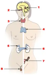
In vertebrates, most hormones belong to the endocrine system, a control system of ductless glands and single cells. In humans, there are eight main glands that generally are considered part of the endocrine system. Other organs of the body also produce and secrete hormones, but are generally not considered part of the endocrine system; these include the heart, kidney, liver, skin, and placenta. The endocrine system works in close relation with the nervous system, and, as noted above, neurohormones are produced by specialized neurons.
Systems involving hormones are so complex and finely tuned that some have speculated that it is irreducibly complex‚ÄĒthat the system could not have evolved over time (and certainly not through the non-purposeful, non-progressive agency of natural selection) because all parts would have had to exist at the same time. However, other scientists have reported findings tracing the evolution of vertebrate steroid hormones from hundreds of millions of years ago, suggesting scenarios for such an evolution by common descent (Bridgham et al. 2006).
Pollution represents a potentially serious problem. Some present-day chemical pollutants, such as PVC, can interfere with hormones. Humanity has a responsibility to carefully manage actions that might negatively impact the environment and disrupt systems that have been developed over many millions of years.
Signaling
Types of signaling
In animals, there are three types of signaling by secreted molecules‚ÄĒendocrine, paracrine, and autocrine‚ÄĒbased on the distance over which the signal acts.
Hormones belong to the first type: They act on target cells distant from their site of synthesis by cells of the endocrine organs. In animals, an endocrine hormone is usually carried by the blood from its site of release to the target cell.
Paracrine signaling molecules only affect target cells in close proximity (an example is the conduction of an impulse), while autocrine cells respond to substances that they themselves release.
However, the designations above are not so clear-cut, as some compounds can participate in two or even three types of signaling. For example, certain small peptides (called neurohormones) function both as neurotransmitters (paracrine signaling) and as hormones (endocrine signaling).
How hormones transmit signals
Hormonal signaling typically involves the following six steps:
- Biosynthesis of the hormone in a specialized tissue.
- Storage and secretion of the hormone.
- Transport of the hormone to the target cell(s), often via the bloodstream.
- Recognition of the hormone by an associated cell membrane or intracellular receptor protein.
- Relay and amplification of the received hormonal signal via a signal transduction process.
- Removal of the signal, which often involves degradation of the hormone, to terminate the cellular response.
Once the hormone reaches the target cell, it binds to, or ‚Äúfits,‚ÄĚ a site on the receptor protein. Binding creates a ligand-receptor complex, causing a conformational change (a change in the molecule's structural arrangement) that ultimately leads to a change in cellular function.
Different cells respond differently to the same ligand. In addition, different ligand-receptor complexes can induce the same biochemical response in some cell types. For example, the hormones glucagon and epinephrine both stimulate increased glucose breakdown in liver cells.
Some hormones bind to receptors embedded in the plasma membrane at the surface of the cell, while others are able to interact with receptors inside the cell (either in the nucleus or the cytoplasm). The former require the aid of molecules called second messengers, such as cyclic AMP, which convey the signal within the cell.
Major classes of vertebrate hormones and their function
Vertebrate hormones may be classified by their chemical make-up. Alternatively, they may be grouped by their solubility and mode of action (i.e., whether they bind to intracellular receptors or to receptors on the cell surface).
According to this latter schema, there are three categories of vertebrate hormones:
- Small lipophilic (lipid-soluble) molecules that are able to diffuse across the plasma membrane of the target cell and interact with intracellular receptors of the cytoplasm or nucleus. The resulting complexes bind to transcription-control regions in DNA, affecting expression of specific genes. The steroid hormones and thyroxine are two examples of this type.
- Lipophilic molecules that bind to cell-surface receptors, such as the eicosanoids.
- Hydrophilic (water-soluble) molecules that bind to receptors on the cell surface because they cannot diffuse across cell membrane. There are two subgroups: (a) peptide hormones, such as insulin, growth hormone, and glucagon, which range in size from a few amino acids to protein-size compounds; and (b) small charged molecules, such as epinephrine and histamine, derived from amino acids, which function as both hormones and neurotransmitters.
Lipophilic molecules that diffuse across the plasma membrane
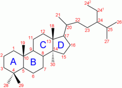
Cholesterol is an important precursor of the steroid hormones, which produce their physiological effects by binding to steroid hormone receptor proteins inside the cytoplasm of the cell. The combined hormone-receptor complex then moves into the nucleus of the cell, where it binds to specific DNA sequences, causing changes in gene transcription and cell function (Beato 1996). It has been shown, however, that some steroid receptors are membrane-associated rather than intracellular (Hamme, 2003).
The five major classes of steroids are as follows:
- Androgens (such as testosterone) are responsible for the development of male secondary sex characteristics.
- Glucocorticoids enable animals to respond to stress. They regulate many aspects of metabolism and immune function, and are often prescribed by doctors to reduce inflammatory conditions like asthma and arthritis.
- Mineralocorticoids help maintain blood volume and control renal excretion of electrolytes.
- Estrogens and progestagens are two classes of sex steroids, a subset of the hormones that produce sex differences or support reproduction.
Thyroxine, produced by thyroid cells, also binds to internal receptors. Thyroid hormones stimulate the breakdown of glucose, fats, and proteins by increasing the levels of many enzymes that catalyze these metabolic reactions.
Lipophilic molecules that bind to cell-surface receptors
Eicosanoids are 20-carbon fatty acids derived from arachidonic acid; the group includes prostaglandins, prostacyclins, thromboxanes, and leukotrienes. Eicosanoids are considered local hormones because they are short-lived; they alter the activities in cells where they are synthesized (autocrine signaling) and in nearby cells (paracrine signaling). Prostaglandins may stimulate inflammation, regulate blood flow, control transport, and induce sleep. Aspirin, for example, works as an anti-inflammatory agent by inhibiting the synthesis of prostaglandin.
Hydrophilic molecules that bind to cell-surface receptors
- Peptide hormones are composed of amino-acid chains. Examples of small peptide hormones are TRH and vasopressin. Peptides composed of scores or hundreds of amino acids, such as insulin and growth hormone, are referred to as protein hormones. More complex protein hormones bear carbohydrate side chains and are called glycoprotein hormones. Luteinizing hormone, follicle-stimulating hormone, and thyroid-stimulating hormone are glycoprotein hormones.

- Some water-soluble hormones are derived from a single amino acid. Histamine, a hormone and neurotransmitter derived from the amino acid histidine, is involved in the dilation of blood vessels. The catecholamines, chemical compounds derived from the amino acid tyrosine, may act as neurohormones. The most abundant catecholamines are epinephrine (adrenaline), norepinephrine (noradrenaline), and dopamine. Released by the adrenal glands in situations of stress, catecholamines cause general physiological changes that prepare the body for physical activity (the fight-or-flight response). Some typical effects are increases in heart rate, blood pressure, blood glucose levels, and a general reaction of the sympathetic nervous system.
The table below provides some examples of water-soluble hormones that bind to cell-surface receptors. The size of the hormone is given in amino acids (note that some hormones have two polypeptide chains of varying lengths, which are designated either A and B or alpha and beta).
| Type | Name | Size | Origin | Major effects |
| Peptide | Follicle-stimulating hormone (FSH) | alpha: 92, beta: 118 | anterior pituitary | Stimulates growth of oocytes and ovarian follices |
| Peptide | Glucagon | 29 | pancreas alpha cells | Stimulates glucose synthesis |
| Peptide | Insulin | A: 21, B: 30 | pancreas beta cells | Regulates glucose uptake; stimulates cell proliferation |
| Peptide | Luteinizing hormone (LH) | 10, beta chain 115 | anterior pituitary | maturation of oocyte; stimulates estrogen and progesterone secretion by ovarian follices |
| Growth factor | nerve growth factor (NGF) | 118 | all tissues innervated by sympathetic neurons | growth and differentiation of sympathetic neurons |
| Growth factor | Epidermal growth factor (EGF) | 53 | salivary and other glands? | Growth of epidermal and other body cells |
| Growth factor | Platelet-derived growth factor | A: 125, B: 109 | platelets and cells in many other tissues | Proliferation of fibroblasts and other cell types; wound healing |
| Neurohormone | Oxytocin | 9 | posterior pituitary gland | Stimulation of smooth muscle contraction |
| Neurohormone | Vasopressin | 9 | posterior pituitary gland | Stimulation of water reabsorption in the kidney |
Regulation
By rate of synthesis
Organisms must be able to respond instantly to many changes in their internal or external environment; such rapid responses are mediated primarily by peptide hormones and catecholamines. Signaling cells that produce them store these hormones in secretory vesicles just under the plasma membrane. All peptide hormones, including [[insulin], are synthesized as part of a longer propolypeptide, which is cleaved (split) by specific enzymes to generate the active molecule just after it is transported to a secretory vesicle. Because of their hydrophilic (water-loving) nature, peptide hormones travel freely in the blood as they dissolve. Peptide hormones mediate short responses that are terminated by their own breakdown.
In contrast, steroid-producing cells, like those in the adrenal cortex, store only a small supply of hormone precursor; when stimulated, they are converted to active hormone, which then diffuses across the cell membrane into the blood. Because cells store little of the active hormone, release takes from hours to days. Steroids are hydrophobic (water-fearing), so they are transported by carrier proteins, and are not rapidly degraded. Thus, responses to thyroxine and steroid hormones take awhile to occur but effects last much longer than those triggered by peptide hormones.
By feedback control
The rate of hormone biosynthesis and secretion is often regulated by feedback circuits, in which changes in the level of one hormone affect the levels of other hormones. This type of control is especially important in coordinating the complex processes of cell growth and differentiation.
By other hormones
The trophic hormones are a special class of hormones that stimulate the hormonal production of endocrine glands. For example, thyroid-stimulating hormone (TSH) causes growth and increased activity of the thyroid, which in turn increases output of thyroid hormones.
Plant hormones
Plant hormones are internally secreted molecules that typically coordinate the responses of plant tissues to environmental signals, such as light or infection.
Plant hormones are traditionally divided into five major groups, although several additional plant hormones have recently been discovered:
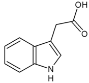
- Auxin was the first plant hormone to be identified; early experiments leading to its discovery were conducted by Charles Darwin in the 1880s. Auxins regulate various aspects of plant development, including cell division and differentiation.
- Abscisic acid (ABA) was originally thought to play a major role in the abscission (shedding) of fruits and in bud dormancy. Some of its confirmed effects (which are mostly inhibitory) include stimulating the closure of stomata in dry conditions, inhibiting shoot growth, and inducing seeds to synthesize proteins.
- Cytokinins (CKs) are active in promoting cell division, and are also involved in cell growth and differentiation.
- Ethylene acts at trace levels throughout the life of the plant by stimulating or regulating the ripening of fruit, the opening of flowers, and the shedding of leaves.
- Gibberellins (GA) are involved in promoting stem elongation and mobilizing food reserves in seeds. The absence of gibberellins results in the dwarfism of some plant varieties.
Non-traditional plant hormones include brassinolide, a plant-specific steroid hormone involved in developmental processes.
The role of hormones in pharmacology
Many hormones and their analogues are used as medication:
- The most commonly prescribed hormones are estrogens and progestagens (as methods of hormonal contraception and as hormone replacement therapy); other steroids (for autoimmune diseases and several respiratory disorders); and thyroxine (as levothyroxine, for hypothyroidism).
- Insulin is used by many diabetics.
- Local preparations for use in otolaryngology (the diagnosis and treatment of ear, nose, and throat disorders) often contain pharmacological equivalents of adrenaline.
- Steroid and vitamin D creams are used extensively in dermatological practice.
A "pharmacological dose" of a hormone is a medical usage referring to an amount of a hormone far greater than its natural occurrence in a healthy body. The effects of pharmacological doses of hormones may be different from responses to naturally occurring amounts and may be therapeutically useful. An example is the ability of pharmacological doses of glucocorticoid to suppress inflammation.
Table of important human hormones
Spelling is not uniform for many hormones. For example, current North American and international usage is estrogen and gonadotropin, while British usage retains the Greek diphthong in oestrogen and the unvoiced aspirant h in gonadotrophin.
| Structure | Name | Abbreviation | Tissue | Cells | Mechanism |
| amine - tryptophan | Melatonin (N-acetyl-5-methoxytryptamine) | pineal gland | pinealocyte | ||
| amine - tryptophan | Serotonin | 5-HT | CNS, GI tract | enterochromaffin cell | |
| amine - tyrosine | Thyroxine (thyroid hormone) | T4 | thyroid gland | thyroid epithelial cell | direct |
| amine - tyrosine | Triiodothyronine (thyroid hormone) | T3 | thyroid gland | thyroid epithelial cell | direct |
| amine - tyrosine (cat) | Epinephrine (or adrenaline) | EPI | adrenal medulla | chromaffin cell | |
| amine - tyrosine (cat) | Norepinephrine (or noradrenaline) | NRE | adrenal medulla | chromaffin cell | |
| amine - tyrosine (cat) | Dopamine | DPM | hypothalamus | ||
| peptide | Antimullerian hormone (or mullerian inhibiting factor or hormone) | AMH | testes | Sertoli cell | |
| peptide | Adiponectin | Acrp30 | adipose tissue | ||
| peptide | Adrenocorticotropic hormone (or corticotropin) | ACTH | anterior pituitary | corticotrope | cAMP |
| peptide | Angiotensinogen and angiotensin | AGT | liver | IP3 | |
| peptide | Antidiuretic hormone (or vasopressin, arginine vasopressin) | ADH | posterior pituitary | varies | |
| peptide | Atrial-natriuretic peptide (or atriopeptin) | ANP | heart | cGMP | |
| peptide | Calcitonin | CT | thyroid gland | parafollicular cell | cAMP |
| peptide | Cholecystokinin | CCK | duodenum | ||
| peptide | Corticotropin-releasing hormone | CRH | hypothalamus | cAMP | |
| peptide | Erythropoietin | EPO | kidney | ||
| peptide | Follicle-stimulating hormone | FSH | anterior pituitary | gonadotrope | cAMP |
| peptide | Gastrin | GRP | stomach, duodenum | G cell | |
| peptide | Ghrelin | stomach | P/D1 cell | ||
| peptide | Glucagon | GCG | pancreas | alpha cells | cAMP |
| peptide | Gonadotropin-releasing hormone | GnRH | hypothalamus | IP3 | |
| peptide | Growth hormone-releasing hormone | GHRH | hypothalamus | IP3 | |
| peptide | Human chorionic gonadotropin | hCG | placenta | syncytiotrophoblast cells | cAMP |
| peptide | Human placental lactogen | HPL | placenta | ||
| peptide | Growth hormone | GH or hGH | anterior pituitary | somatotropes | |
| peptide | Inhibin | testes | Sertoli cells | ||
| peptide | Insulin | INS | pancreas | beta cells | tyrosine kinase |
| peptide | Insulin-like growth factor (or somatomedin) | IGF | liver | tyrosine kinase | |
| peptide | Leptin | LEP | adipose tissue | ||
| peptide | Luteinizing hormone | LH | anterior pituitary | gonadotropes | cAMP |
| peptide | Melanocyte stimulating hormone | MSH or őĪ-MSH | anterior pituitary/pars intermedia | cAMP | |
| peptide | Oxytocin | OXT | posterior pituitary | IP3 | |
| peptide | Parathyroid hormone | PTH | parathyroid gland | parathyroid chief cell | cAMP |
| peptide | Prolactin | PRL | anterior pituitary | lactotrophs | |
| peptide | Relaxin | RLN | varies | ||
| peptide | Secretin | SCT | duodenum | S cell | |
| peptide | Somatostatin | SRIF | hypothalamus, islets of Langerhans | delta cells | |
| peptide | Thrombopoietin | TPO | liver, kidney | ||
| peptide | Thyroid-stimulating hormone | TSH | anterior pituitary | thyrotropes | cAMP |
| peptide | Thyrotropin-releasing hormone | TRH | hypothalamus | IP3 | |
| steroid - glu. | Cortisol | adrenal cortex (zona fasciculata) | direct | ||
| steroid - min. | Aldosterone | adrenal cortex (zona glomerulosa) | direct | ||
| steroid - sex (and) | Testosterone | testes | Leydig cells | direct | |
| steroid - sex (and) | Dehydroepiandrosterone | DHEA | multiple | direct | |
| steroid - sex (and) | Androstenedione | adrenal glands, gonads | direct | ||
| steroid - sex (and) | Dihydrotestosterone | DHT | multiple | direct | |
| steroid - sex (est) | Estradiol | E2 | ovary | granulosa cells | direct |
| steroid - sex (est) | Estrone | ovary | granulosa cells | direct | |
| steroid - sex (est) | Estriol | placenta | syncytiotrophoblast | direct | |
| steroid - sex (pro) | Progesterone | ovary, adrenal glands, placenta | granulosa cells | direct | |
| sterol | Calcitriol (Vitamin D3) | skin/proximal tubule of kidneys | direct | ||
| eicosanoid | Prostaglandins | PG | seminal vesicle | ||
| eicosanoid | Leukotrienes | LT | white blood cells | ||
| eicosanoid | Prostacyclin | PGI2 | endothelium | ||
| eicosanoid | Thromboxane | TXA2 | platelets |
Code:
- (and) = "Androgen is the generic term for any natural or synthetic compound, usually a steroid hormone, that stimulates or controls the development and maintenance of masculine characteristics in vertebrates by binding to androgen receptors."
- (cat) = "Catecholamines are chemical compounds derived from the amino acid tyrosine containing catechol and amine groups."
- (est) = "Estorgen, a group of steroid compounds, named for their importance in the estrous cycle, and functioning as the primary female sex hormone."
- (pro) = "Progestagens (also spelled progestogens or gestagens) are hormones which produce effects similar to progesterone, the only natural progestagen."
ReferencesISBN links support NWE through referral fees
- Beato, M., S. Chavez, and M. Truss. 1996. Transcriptional regulation by steroid hormones. Steroids 61(4): 240-251.
- Bridgham, J. T., S. M. Carroll, and J. W. Thornton. 2006. Evolution of Hormone-Receptor Complexity by Molecular Exploitation. Science 312: 97-101.
- Cooper, G. M., and R. E. Hausman. 2004. The Cell: A Molecular Approach. Washington, D.C.: ASM Press. ISBN 0878932143
- Hammes, S. R. 2003. The further redefining of steroid-mediated signaling. Proc Natl Acad Sci 100(5): 2168-70.
- Lodish, H., D. Baltimore, A. Berk, S. L. Zipursky, P. Matsudaira, and J. Darnell. 1995. Molecular Cell Biology. New York: Scientific American Books. ISBN 0716723808
- Mathews, C. K. and K. E. van Holde. 1990. Biochemistry. San Francisco: Benjamin-Cummings. ISBN 0805350152
- Stryer, L. 1995. Biochemistry, New York: W.H. Freeman. ISBN 0716720094
| Hormones and endocrine glands - edit |
|---|
|
Hypothalamus: GnRH - TRH - CRH - GHRH - somatostatin - dopamine | Posterior pituitary: vasopressin - oxytocin | Anterior pituitary: GH - ACTH - TSH - LH - FSH - prolactin - MSH - endorphins - lipotropin Thyroid: T3 and T4 - calcitonin | Parathyroid: PTH | Adrenal medulla: epinephrine - norepinephrine | Adrenal cortex: aldosterone - cortisol - DHEA | Pancreas: glucagon- insulin - somatostatin | Ovary: estradiol - progesterone - inhibin - activin | Testis: testosterone - AMH - inhibin | Pineal gland: melatonin | Kidney: renin - EPO - calcitriol - prostaglandin | Heart atrium: ANP Stomach: gastrin | Duodenum: CCK - GIP - secretin - motilin - VIP | Ileum: enteroglucagon | Liver: IGF-1 Placenta: hCG - HPL - estrogen - progesterone Adipose tissue: leptin, adiponectin Target-derived NGF, BDNF, NT-3 |
Credits
New World Encyclopedia writers and editors rewrote and completed the Wikipedia article in accordance with New World Encyclopedia standards. This article abides by terms of the Creative Commons CC-by-sa 3.0 License (CC-by-sa), which may be used and disseminated with proper attribution. Credit is due under the terms of this license that can reference both the New World Encyclopedia contributors and the selfless volunteer contributors of the Wikimedia Foundation. To cite this article click here for a list of acceptable citing formats.The history of earlier contributions by wikipedians is accessible to researchers here:
- Hormone  history
- Catecholamine  history
- Androgen  history
- Estrogen  history
- Progestagen  history
- Plant_hormone  history
- Thyroxine  history
- Histamine  history
The history of this article since it was imported to New World Encyclopedia:
Note: Some restrictions may apply to use of individual images which are separately licensed.


