Oxytocin

| |
Oxytocin
| |
| Systematic name | |
| IUPAC name  ? | |
| Identifiers | |
| CAS number | 50-56-6 |
| ATC code | H01BB02 |
| PubChem | 439302 |
| DrugBank | BTD00016 |
| Chemical data | |
| Formula | C43H66N12O12S2  |
| Mol. weight | 1007.19 g/mol |
| Pharmacokinetic data | |
| Bioavailability | nil |
| Protein binding | 30% |
| Metabolism | hepatic oxytocinases |
| Half life | 1-6 min |
| Excretion | Biliary and renal |
| Therapeutic considerations | |
| Pregnancy cat. | ? |
| Legal status | ? |
| Routes | Intranasal, IV, IM |
Oxytocin (ŇŹk'sń≠-tŇć'sń≠n) is a relatively small polypeptide hormone in mammals that plays an important role in birth and ejection of milk from the female breast. It also acts as a neurotransmitter in the brain. Along with the antidiuretic hormone vassopressin, oxytocin is one of the two major hormones released from the posterior lobe of the pituitary gland (Blakemore and Jennett 2001).
Ocytocin, which means "quick birth" in Greek, is released in large amounts in females after distension of the cervix and vagina during labor, stimulating smooth muscle contractions of the uterus and facilitating childbirth. It also is released after stimulation of the nipples, inducing muscular contractions around the alveoli and milk ducts in the breasts, facilitating breastfeeding.
In humans, oxytocin is involved in social recognition and bonding, and might be involved in the formation of trust between people (Kosfeld 2005). Also, oxytocin has been known to affect the brain by regulating circadian homeostasis, such as a person's body temperature, activity level, and wakefulness (Kraft 2007). In humans, oxytocin is released during orgasm in both sexes.
Oxytocin involves harmonious interaction between neural and hormonal systems. It is produced in nerve cells rather than in glandular cells (where most hormones are made) and it is released into the blood following sensory nerve stimulation of the nerve cells (Blakemore and Jennett 2001). For example, the suckling, sight, and sound of an infant, among other stimuli associated with breastfeeding, stimulate communication with hypothalamic nerve cells (where the hormone is produced). This leads to secretion of the hormone from the pituitary gland, where the ending of the nerves lie and the hormone is packaged in vesicles (Blakemore and Jennett 2001). The action of oxytocin occurs relatively rapidly because sensory nerve impulses are involved.
oxytocin, prepro- (neurophysin I)
| |
| Identifiers | |
| Symbol | OXT |
| Alt. Symbols | OT |
| Entrez | 5020 |
| HUGO | 8528 |
| OMIM | 167050 |
| RefSeq | NM_000915 |
| UniProt | P01178 |
| Other data | |
| Locus | Chr. 20 p13 |
Structure
Ocytocin is a hormone, meaning it is a chemical messenger secreted by cells (including tissues and organs) in one part of a multicellular organism to travel to and coordinate the activities of different cells, providing a value to the whole organism. An enormous range of chemicals are used for this type of cell-to-cell communication, including peptides (chains of amino acids) and steroids (a type of fat-soluble organic compound). Oxytocin is a peptide hormone.
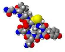
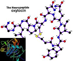
Oxytocin has the chemical formula C43H66N12O12S2. It is a relatively short polypeptide, being composed of only nine amino acids (a nonapeptide). The sequence is cysteine - tyrosine - isoleucine - glutamine - asparagine - cysteine - proline - leucine - glycine (CYIQNCPLG). The cysteine residues form a sulfur bridge. Oxytocin has a molecular mass of 1007 daltons. One international unit (IU) of oxytocin is the equivalent of about two micrograms of pure peptide.
The structure of oxytocin is very similar to that of vasopressin, an antidiuretic hormone that is also a nonapeptide: cysteine - tyrosine - phenylalanine - glutamine - asparagine - cysteine - proline - arginine - glycine). Vassopressin, whose residues also form a sulfur bridge, has a sequence that differs from oxytocin by two amino acids.
Oxytocin and vasopressin are the only known hormones released by the human posterior pituitary gland to act at a distance. However, oxytocin neurons make other peptides, including corticotropin-releasing hormone (CRH) and dynorphin, for example, that act locally. The magnocellular neurons that make oxytocin are adjacent to magnocellular neurons that make vasopressin, and are similar in many respects.
Oxytocin was the first hormone for which the structure was identified and which was synthesized in the laboratory (Blakemore and Jennett 2001). Oxytocin and vasopressin were isolated and synthesized by Vincent du Vigneaud in 1953, work for which he received the Nobel Prize in Chemistry in 1955.
Synthesis, storage and release
Oxytocin is made in magnocellular neurosecretory cells in the supraoptic nucleus and paraventricular nucleus of the hypothalamus and is released into the blood from the posterior lobe of the pituitary gland.
The posterior pituitary essentially contains the endings of nerves whose cell bodies lie in the hypothalamus (Blakemore and Jennett 2001). The hormone is manufactured in the cell bodies in the hypothalamus in the form of a larger, precursor molecule. It is then transported down the nerve fibers to the posterior lobe, where the active hormone is cleaved from the precursor molecule and then is secreted directly into the blood capillaries from the nerve endings of the posterior pituitary (Blakemore and Jennett 2001).
In the pituitary gland, oxytocin is packaged in large, dense-core vesicles, where it is bound to neurophysin I; neurophysin is a large peptide fragment of the giant precursor protein molecule from which oxytocin is derived by enzymatic cleavage.
Secretion of oxytocin from the neurosecretory nerve endings is regulated by the electrical activity of the oxytocin cells in the hypothalamus. These cells generate action potentials that propagate down axons to the nerve endings in the pituitary; the endings contain large numbers of oxytocin-containing vesicles, which are released by exocytosis when the nerve terminals are depolarized.
Oxytocin also is made by some neurons in the paraventricular nucleus that project to other parts of the brain and to the spinal cord.
Virtually all vertebrates have an oxytocin-like nonapeptide hormone that supports reproductive functions and a vasopressin-like nonapeptide hormone involved in water regulation. The two genes are always located close to each other (less than 15,000 bases apart) on the same chromosome and are transcribed in opposite directions. It is thought that the two genes resulted from a gene duplication event; the ancestral gene is estimated to be about 500 million years old and is found in cyclostomes (modern members of the Agnatha) (Gimpl and Fahrenholz 2001).
Actions
Oxytocin has peripheral (hormonal) actions, and also has actions in the brain. The actions of oxytocin are mediated by specific, high-affinity oxytocin receptors. The oxytocin receptor is a G-protein-coupled receptor, which requires Mg2+ and cholesterol. It belongs to the rhodopsin-type (class I) group of G-protein-coupled receptors.
Peripheral (hormonal) actions
The peripheral actions of oxytocin mainly reflect secretion from the pituitary gland.
- Letdown reflect. In lactating (breastfeeding) mothers, oxytocin acts at the mammary glands, causing milk to be "let down" into a collecting chamber, from where it can be extracted by sucking at the nipple. Sucking by the infant at the nipple is relayed by spinal nerves to the hypothalamus. The stimulation causes neurons that make oxytocin to fire action potentials in intermittent bursts; these bursts result in the secretion of pulses of oxytocin from the neurosecretory nerve terminals of the pituitary gland.
- Uterine contraction. Uterine contraction is important for cervical dilation before birth and causes contractions during the second and third stages of labor. Also, oxytocin release during breastfeeding causes mild but often painful uterine contractions during the first few weeks of lactation. This also serves to assist the uterus in clotting the placental attachment point postpartum. However, in knockout mice lacking the oxytocin receptor, reproductive behavior and parturition is normal (Takayanagi 2005).
- Orgasm and sperm transport. Oxytocin is secreted into the blood at orgasm in both males and females (Carmichael et al. 1987). In males, oxytocin may facilitate sperm transport in ejaculation.
- Urine and sodium excretion. Due to its similarity to vasopressin, oxytocin can reduce the excretion of urine slightly. More important, in several species, oxytocin can stimulate sodium excretion from the kidneys (natriuresis), and in humans, high doses of oxytocin can result in hyponatremia.
- Possible embryonal development in rodents. Oxytocin and oxytocin receptors are also found in the heart in some rodents, and the hormone may play a role in the embryonal development of the heart by promoting cardiomyocyte differentiation (Paquin et al. 2002; Jankowski et al. 2004). However, the absence of either oxytocin or its receptor in knockout mice has not been reported to produce cardiac insufficiencies (Takayanagi 2005).
Actions of oxytocin within the brain
Oxytocin secreted from the pituitary gland cannot re-enter the brain because of the blood-brain barrier. Instead, the behavioral effects of oxytocin are thought to reflect release from centrally projecting oxytocin neurons, different from those that project to the pituitary gland. Oxytocin receptors are expressed by neurons in many parts of the brain and spinal cord, including the amygdala, ventromedial hypothalamus, septum, and brainstem.
- Sexual arousal. Oxytocin injected into the cerebrospinal fluid causes spontaneous erections in rats (Gimpl and Fahrenholz 2001), reflecting actions in the hypothalamus and spinal cord.
- Bonding. In the prairie vole, oxytocin released into the brain of the female during sexual activity is important for forming a monogamous pair bond with her sexual partner. Vasopressin appears to have a similar effect in males (Broadfoot 2002). In people, plasma concentrations of oxytocin have been reported to be higher among people who claim to be falling in love. Oxytocin has a role in social behaviors in many species, and so it seems likely that it has similar roles in humans.
- Autism. A 1998 report on a research study noted significantly lower levels of oxytocin in blood plasma of autistic children (Modahl et al. 1998). In 2003, a research team reported a decrease in autism spectrum repetitive behaviors when oxytocin was administered intravenously (Hallander et al. 2003). A 2007 study reported that oxytocin helped autistic adults retain the ability to evaluate the emotional significance of speech intonation (Hollander et al. 2007).
- Maternal behavior. Sheep and rat females given oxytocin antagonists after giving birth do not exhibit typical maternal behavior. By contrast, virgin female sheep show maternal behavior towards foreign lambs upon cerebrospinal fluid infusion of oxytocin, which they would not do otherwise (Kendrick 2007).
- Increasing trust and reducing fear. In a risky investment game, experimental subjects given nasally administered oxytocin displayed "the highest level of trust" twice as often as the control group. Subjects who were told that they were interacting with a computer showed no such reaction, leading to the conclusion that oxytocin was not merely affecting risk-aversion (Kosfeld et al. 2005). Nasally administered oxytocin has also been reported to reduce fear, possibly by inhibiting the amygdala (which is thought to be responsible for fear responses) (Kirsch et al. 2005). There is no conclusive evidence for passage of oxytocin to the brain through intranasal administration, however.
- Tolerance to drugs. According to some studies in animals, oxytocin inhibits the development of tolerance to various addictive drugs (opiates, cocaine, alcohol) and reduces withdrawal symptoms (Kovacs et al. 1998).
- Preparing fetal neurons for delivery. Crossing the placenta, maternal oxytocin reaches the fetal brain and induces a switch in the action of neurotransmitter GABA from excitatory to inhibitory on fetal cortical neurons. This silences the fetal brain for the period of delivery and reduces its vulnerability to hypoxic damage (Tyzio et al. 2006).
- Learning. Certain learning and memory functions are impaired by centrally administered oxytocin (Gimpl and Fahrenholz 2001).
- MDMA function. The illicit party drug MDMA (ecstasy) may increase feelings of love, empathy, and connection to others by stimulating oxytocin activity via activation of serotonin 5HT1A receptors, if initial studies in animals apply to humans (Thompson et al. 2007).
Drug forms
Synthetic oxytocin is sold as medication under the trade names Pitocin and Syntocinon and also as generic Oxytocin. Oxytocin is destroyed in the gastrointestinal tract, and therefore must be administered by injection or as nasal spray. Oxytocin has a half-life of typically about three minutes in the blood. Oxytocin given intravenously does not enter the brain in significant quantities‚ÄĒit is excluded from the brain by the blood-brain barrier. Drugs administered by nasal spray are thought to have better access to the central nervous system. Oxytocin nasal sprays have been used to stimulate breastfeeding.
Injected oxytocin analogues are used to induce labor and support labor in case of non-progression of parturition. It has largely replaced ergotamine as the principal agent to increase uterine tone in acute postpartum haemorrhage. Oxytocin also is used in veterinary medicine to facilitate birth and to increase milk production. The tocolytic agent atosiban (Tractocile¬ģ) acts as an antagonist of oxytocin receptors; this drug is registered in many countries to suppress premature labor between 24 and 33 weeks of gestation. It has fewer side effects than drugs previously used for this purpose (ritodrine, salbutamol, and terbutaline).
Some have suggested that the trust-inducing property of oxytocin might help those who suffer from social anxieties, while others have noted the potential for abuse by swindlers given the trust associated with oxytocin usage.
Potential adverse reactions
Oxytocin is relatively safe when used at recommended doses. Potential side effects include:
- Central nervous system: Subarachnoid hemorrhage, seizures.
- Cardiovascular: Increased heart rate, blood pressure, systemic venous return, cardiac output, and arrhythmias.
- Genitourinary: Impaired uterine blood flow, pelvic hematoma, tetanic uterine contractions, uterine rupture, postpartum hemorrhage.
ReferencesISBN links support NWE through referral fees
- Blakemore, C., and S. Jennett. 2001. The Oxford Companion to the Body. New York: Oxford University Press. ISBN 019852403X
- Broadfoot, M. V. 2002. High on Fidelity. What can voles teach us about monogamy? American Scientist. Retrieved October 20, 2007.
- Caldwell, H. K., and W. S. Young. 2006. Oxytocin and Vasopressin: Genetics and behavioral implications. In R. Lim and A. Lajtha, eds. Handbook of Neurochemistry and Molecular Neurobiology. 3rd edition. New York: Springer. ISBN 0387303480. Retrieved October 20, 2007.
- Carmichael, M. S., R. Humbert, J. Dixen, G. Palmisano, W. Greenleaf, and J. M. Davidson. 1987. Plasma oxytocin increases in the human sexual response. J. Clin. Endocrinol. Metab. 64:27‚Äď31. PMID 3782434.
- Gimpl, G., and F. Fahrenholz. 2001. The oxytocin receptor system: Structure, function, and regulation. Physiological Reviews 81. PMID 11274341. Retrieved October 20, 2007.
- Hollander, E., S. Novotny, M. Hanratty, et al. 2003. Oxytocin infusion reduces repetitive behaviors in adults with autistic and Asperger's disorders. Neuropsychopharmacology 28(1):193‚Äď198. PMID 12496956. Retrieved October 20, 2007.
- Hollander, E., J. Bartz, W. Chaplin, et al. 2007. Oxytocin increases retention of social cognition in autism. Biol Psychiatry 61(4):498‚Äď503. PMID 16904652.
- Jankowski, M., B. Danalache, D. Wang, et al. 2004. Oxytocin in cardiac ontogeny. Proc. Nat'l. Acad. Sci. USA 101:13074‚Äď13079. PMID 15316117.
- Kendrick, K. M. 2007. The neurobiology of social bonds. Journal of Neuroendocrinology. Retrieved October 20, 2007.
- Kirsch, P., et al. 2005. Oxytocin modulates neural circuitry for social cognition and fear in humans. J. Neurosci. 25:11489‚Äď11493. PMID 16339042.
- Kosfeld, M., et al. 2005. Oxytocin increases trust in humans. Nature 435:673‚Äď676. PMID 15931222. Retrieved October 20, 2007.
- Kovacs, G. L., Z. Sarnyai, and G. Szabo. 1998. Oxytocin and addiction: A review. Psychoneuroendocrinology 23:945‚Äď962. PMID 9924746.
- Kraft, U. 2007. Rhythm and blues. Scientific American June/July 2007. Retrieved October 20, 2007.
- Modahl, C., L. Green, D. Fein, et al. 1998. Plasma oxytocin levels in autistic children. Biol. Psychiatry 43(4):270‚Äď277. PMID 9513736.
- Paquin, J., et al. 2002. Oxytocin induces differentiation of P19 embryonic stem cells to cardiomyocytes. Proc. Nat'l. Acad. Sci. USA 99:9550‚Äď9555. PMID 12093924.
- Takayanagi, Y., et al. 2005. Pervasive social deficits, but normal parturition, in oxytocin receptor-deficient mice. Proc. Nat'l. Acad. Sci. USA 102:16096‚Äď160101. PMID 16249339.
- Thompson, M. R., P. D. Callaghan, G. E. Hunt, J. L. Cornish, and I. S. McGregor. 2007. A role for oxytocin and 5-HT(1A) receptors in the prosocial effects of 3,4 methylenedioxymethamphetamine ("ecstasy"). Neuroscience 146:509‚Äď514. PMID 17383105.
- Tyzio, R., et al. 2006. Maternal oxytocin triggers a transient inhibitory switch in GABA signaling in the fetal brain during delivery. Science 314:1788‚Äď1792. PMID 17170309.
Credits
New World Encyclopedia writers and editors rewrote and completed the Wikipedia article in accordance with New World Encyclopedia standards. This article abides by terms of the Creative Commons CC-by-sa 3.0 License (CC-by-sa), which may be used and disseminated with proper attribution. Credit is due under the terms of this license that can reference both the New World Encyclopedia contributors and the selfless volunteer contributors of the Wikimedia Foundation. To cite this article click here for a list of acceptable citing formats.The history of earlier contributions by wikipedians is accessible to researchers here:
The history of this article since it was imported to New World Encyclopedia:
Note: Some restrictions may apply to use of individual images which are separately licensed.
