Heart

In anatomy, the heart is a muscular, pumping organ of the closed circulatory system of all vertebrates and some invertebrates (annelids and cephalopods) that is responsible for moving blood through the blood vessels by automatic, repeated, rhythmic contractions, or a similar structure for moving blood in the open circulatory system used by some invertebrates (arthropods and some mollusks).
The term cardiac (as in cardiology and cardiovascular system) means "related to the heart" and comes from the Greek καρδία, kardia, for "heart" (AHSMD 2004). Cardiovascular disease refers to the class of diseases that involve the heart and/or blood vessels (arteries, and veins).
The average human heart beating at 72 beats per minute, will beat approximately 2.5 billion times during a lifetime of 66 years.
Overview
The heart is part of a living organism's circulatory system, which is also referred to as a cardiovascular system. The circulatory system is an organ system that moves substances to and from cells; it can also play a part in homeostasis by helping stabilize body temperature and pH. An organ identified as a heart can be found in either an open or a closed circulatory system.
Open circulatory system. An open circulatory system is an arrangement of internal transport in which circulatory fluid, in a cavity called the hemocoel (also spelled haemocoel), bathes the organs directly. There is no distinction between blood and interstitial fluid; this combined fluid is called hemolymph (also spelled haemolymph).
Open systems are present in some invertebrates, like mollusks and arthropods. Muscular movements during locomotion by animals with such a system can facilitate hemolymph movement, but diverting flow from one area to another is limited. When the heart relaxes, blood is drawn back toward the heart through open-ended pores.
Hemolymph fills all of the interior hemocoel of the body and surrounds all cells. Hemolymph is composed of water, inorganic salts (mostly Na+, Cl-, K+, Mg2+, and Ca2+), and organic compounds (mostly carbohydrates, proteins, and lipids). The primary oxygen transporter molecule is hemocyanin.
There are free-floating cells, the hemocytes, within the hemolymph. They play a role in the arthropod immune system.
Closed circulatory system
The circulatory systems of all vertebrates, annelids (for example, earthworms), and cephalopods (squid and octopus) are closed, meaning that the blood never leaves the system of blood vessels, which consists of arteries, veins, and capillaries.
The main components of the closed circulatory system are the heart, the blood, and the blood vessels. Arteries bring oxygenated blood to the tissues (except pulmonary arteries), and veins bring deoxygenated blood back to the heart (except pulmonary veins). Blood passes from arteries to veins through capillaries, which are the thinnest and most numerous of the blood vessels.
The closed circulatory systems of fish, amphibians, reptiles, birds, and mammals show various stages of sophistication.
In fish, the system has only one circuit, with the blood being pumped through the capillaries of the gills and on to the capillaries of the body tissues. This is known as single circulation. The heart of fish is therefore only a single pump (consisting of two chambers).
In amphibians and most reptiles, a double circulatory system is used, but the heart is not always completely separated into two pumps. Amphibians have a three-chambered heart.
Birds and mammals show complete separation of the heart into two pumps, for a total of four heart chambers; it is thought that the four-chambered heart of birds evolved independently of that of mammals.
Early development

It is unknown how blood in the embryo circulates for the first 21 days in the absence of a functioning heart, although some have hypothesized that the heart is not so much a pump, as a Hydraulic Ram — an organ built-up from cumulative peripheral activity. [1]
When the human embryonic heart begins beating — around 21 days after conception, or five weeks after the last normal menstrual period (LMP), which is the date normally used to date pregnancy. The human heart begins beating at a rate near the mother’s, about 75-80 beats per minute (BPM). The embryonic heart rate (EHR) then accelerates linearly for the first month of beating, peaking at 165-185 BPM during the early 7th week, (early 9th week after the LMP). This acceleration is approximately 3.3 BPM per day, or about 10 BPM every three days, an increase of 100 BPM in the first month.[1]
After peaking at about 9.2 weeks after the LMP, it decelerates to about 152 BPM (+/-25 BPM) during the 15th week after the LMP. After the 15th week the deceleration slows reaching an average rate of about 145 (+/-25 BPM) BPM at term. The regression formula which describes this acceleration before the embryo reaches 25 mm in crown-rump length or 9.2 LMP weeks is Age in days = EHR(0.3)+6
There is no difference in male and female heart rates before birth.[2]
Structure
In the human body, the heart is usually situated in the middle of the thorax with the largest part of the heart slightly offset to the left (although sometimes it is on the right, see dextrocardia), underneath the breastbone (see diagrams). The heart is usually felt to be on the left side because the left heart (left ventricle) is stronger (it pumps to all body parts). The left lung is smaller than the right lung because the heart occupies more of the left hemithorax. The heart is enclosed by a sac known as the pericardium and is surrounded by the lungs. The pericardium comprises two parts: the fibrous pericardium, made of dense fibrous connective tissue; and a double membrane structure containing a serous fluid to reduce friction during heart contractions (the serous pericardium). The mediastinum, a subdivision of the thoracic cavity, is the name of the heart cavity.
The apex is the blunt point situated in an inferior (pointing down and left) direction. A stethoscope can be placed directly over the apex so that the beats can be counted. It is located posterior to the 5th intercostal space in the left mid-clavicular line. In normal adults, the mass of the heart is 250-350 g (9-12 oz), or about three quarters the size of a clenched fist, but extremely diseased hearts can be up to 1000 g (2 lb) in mass due to hypertrophy. It consists of four chambers, the two upper atria (singular: atrium ) and the two lower ventricles. On the left is a picture of a fresh human heart which was removed from a 64-year-old male.
The heart is composed of cardiac muscle, an involuntary muscle tissue of the autonomic nervous system, which is found only within this organ (AHSMD 2004). .[3]
Functioning

The function of the right side of the heart (see right heart) is to collect de-oxygenated blood, in the right atrium, from the body and pump it, via the right ventricle, into the lungs (pulmonary circulation) so that carbon dioxide can be dropped off and oxygen picked up (gas exchange). This happens through the passive process of diffusion. The left side (see left heart) collects oxygenated blood from the lungs into the left atrium. From the left atrium the blood moves to the left ventricle which pumps it out to the body. On both sides, the lower ventricles are thicker and stronger than the upper atria. The muscle wall surrounding the left ventricle is thicker than the wall surrounding the right ventricle due to the higher force needed to pump the blood through the systemic circulation.
Starting in the right atrium, the blood flows through the tricuspid valve to the right ventricle. Here it is pumped out the pulmonary semilunar valve and travels through the pulmonary artery to the lungs. From there, blood flows back through the pulmonary vein to the left atrium. It then travels through the bicuspid valve to the left ventricle, from where it is pumped through the aortic semilunar valve to the aorta. The aorta forks, and the blood is divided between major arteries which supply the upper and lower body. The blood travels in the arteries to the smaller arterioles, then finally to the tiny capillaries which feed each cell. The (relatively) deoxygenated blood then travels to the venules, which coalesce into veins, then to the inferior and superior venae cavae and finally back to the right atrium where the process began.
The heart is effectively a syncytium, a meshwork of cardiac muscle cells interconnected by contiguous cytoplasmic bridges. This relates to electrical stimulation of one cell spreading to neighboring cells. Its pumping is so strong, that if the aorta would be cut off, then the blood would squirt to a level of 4 meters [4].
First aid
See cardiac arrest for emergencies involving the heart
If a person is encountered in cardiac arrest (no heartbeat), cardiopulmonary resuscitation (CPR) should be started and help called. If an automated external defibrillator is available, this device may automatically administer defibrillation if this is indicated. Usually, if there is enough time, the person can be rushed to the hospital where he or she will be cared for by a cardiologist, a doctor who specializes in the heart and lungs.
Electrical innervation of the heart in health is supplied by two closely intertwined mechanisms. The first mechanism is well demonstrated in electrical coil systole (interpreted by the electrocardiogram as QRS)as an individualized myocardial electrical tree initiated by the sinoatrial node. Secondary diastolic electrical control is posited to represent autonomic recoil control from the vagus nerve and cardiac branches and the thoracic ganglia.
Food use
The hearts of cattle, sheep, pigs, chickens and certain fowl are consumed in many countries. They are counted among offal, but being a muscle, the taste of heart is like regular meat. It resembles venison in structure and taste.
Aztec heart removal
The Aztec civilization used the heart as a sacrificial token during the sacrifice of a human being. The priest used a stone knife to cut into the thoracic cavity and remove the heart, upon which it would be placed on a stone altar as an offering to the gods. The greatest sacrifice under the reign of Montezuma involved the removal of the hearts of over 12,000 enemy soldiers.
"Heart" as a symbol

The heart (♥) has long been used as a symbol to refer to the spiritual, emotional, moral, and in the past also intellectual core of a human being. As the heart was once widely believed to be the seat of the human mind, the word heart continues to be used poetically to refer to the soul, and stylized depictions of hearts are extremely prevalent symbols representing love. However, more realistic depictions of human hearts tend to have macabre connotations of death and violence, quite unlike the concepts associated with the poetic and symbolic heart. This discrepancy is a common source of dark humor.
As metaphor
In mythology, spirituality and religion
In religious texts such as the Bible [2], the heart has historically been ascribed much mystical significance, either as metaphor or as an organ genuinely believed to have spiritual or divine attributes.
In the Bible, this idea emerges in the earliest passages; Genesis 6:5 situates the thoughts of evil men in their hearts, and Exodus 5 through 12 speak repeatedly of the Lord "hardening Pharaoh's heart." By this it is meant that God made Pharaoh resolve not to let the Israelite slaves leave Egypt, in order to bring judgment against Pharaoh and demonstrate his power: "'Go to Pharaoh, for I have hardened his heart and the hearts of his officials so that I may perform these miraculous signs of mine among them'" (Exodus 10:1). In the Book of Jeremiah 17:9, it is written that the "heart is deceitful above all things, and desperately wicked," and that the Lord is the judge who "tries" the human heart.
The Sacred Heart of Jesus and the Immaculate Heart of Mary are traditional Roman Catholic devotional images.
Similarly, in Egyptian mythology, the heart was weighed in a balance against the feather of Ma'at, symbolising truth, in the judgment of the dead in the Egyptian Book of the Dead. Egyptian sources do not actually reveal whether the heart had to be lighter or heavier than the feather for the deceased to pass into paradise - all depictions show only the weighing of the heart, not the actual results, heavier or lighter. (See also Egyptian soul).
In early science and philosophy
Many classical and medieval philosophers and scientists, including Aristotle, considered the heart the seat of thought, reason or emotion, often rejecting the value of the brain.
The Roman physician Galen located the seat of the passions in the liver, the seat of reason in the brain, and considered the heart to be the seat of the emotions. While Galen's identification of the heart with emotion were proposed as a part of his theory of the circulatory system, the heart has continued to be used as a symbolic source of human emotions even after the rejection of such beliefs.
The Stoics taught that the heart was the seat of the human soul.
As icon
In European traditional art and folklore, the heart symbol is drawn in a stylized shape. This shape is typically colored red, suggesting both blood and, in many cultures, passion and strong emotion. The hearts and the diamonds are the two red suits in most playing card decks. The shape is particularly associated with romantic poetry; it is often seen on St. Valentine's Day cards, candy boxes, and similar popular culture artifacts as a symbol of romantic love.
What the traditional "heart shape" actually depicts is a matter of some controversy. It only vaguely resembles the human heart. Some people claim that it actually depicts the heart of a cow, a more readily available sight to most people in past centuries than an actual human heart. However, while bovine hearts are more similar to the iconic heart shape, the resemblance is still slight. The shape does resemble that of the three-chambered heart of the turtle, and that of the human male prostate gland, but it is very unlikely that the image was patterned after either of these organs.
The "heart" shape could also be considered to depict features of the human female body, such as the female's pubic mound or spread vulva. The tantric symbol of the "Yoni" is another example of a heart-shaped abstraction of a woman's vulva. In fact, the symmetry resembles the vulva far more than the asymmetry of the organ. In the introduction to the Vagina Monologues Gloria Steinem writes, "[The heart] was reduced from power to romance by centuries of male dominance."
Others maintain that the heart resembles the shape of the female breasts or the female buttocks.
Another possible origin can be seen on the coins of the ancient city of Cyrene, some of which depict the seeds or fruit of the now-extinct silphium plant. The seeds are distinctly heart-shaped. Since this plant was widely used as an ancient herbal contraceptive or abortifacient, this shape may have come to be associated with sexuality and love.
The "heart" shape is formed by the back and wings of a dove, which was associated with Aphrodite, the ancient Greek goddess of love.
It may also be a stylised form of the combination of the silhouettes of two people kissing.
The most common emoticon for the heart is <3. In Unicode, the heart symbol is U+2665, and it can thus be generated in HTML by typing ♥ or ♥, or by the HTML entity ♥. Mathematically, a heart-shaped figure, called a cardioid, can be represented by plotting a graph of either or, in polar form,


Additional images
- 2cb.jpg
2C-B pill.

Mating Mute Swans may appear to make a negative space heart.
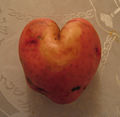
A red potato exhibiting gemination, an effect brought about by a partial twinning during development.

Heart carved in tree


See also
- Cardiac cycle
- Electrocardiogram
- Electrical conduction system of the heart
ReferencesISBN links support NWE through referral fees
- ↑ http://www.obgyn.net/us/us.asp?page=/us/cotm/0001/ehr2000
- ↑ Terry J. DuBose http://www.obgyn.net/english/pubs/features/dubose/ehr-age.htm Sex, Heart Rate and Age]
- ↑ The American Heritage Stedman's Medical Dictionary. KMLE Medical Dictionary Definition of cardiac.
- ↑ Ian Holm in From Hell
References
- The Shape of My Heart: Where did the ubiquitous Valentine's symbol come from? by Keelin McDonell, Slate.com.
- www.heartsymbol.com: The Heart Symbol - Origin, History And Significance by Prof. Armin Dietz
References
The American Heritage® Stedman's Medical Dictionary (AHSMD 2004)., 2nd edition Copyright ⓒ 2004, 2002, 2001, 1995 by Houghton Mifflin Company. Published by Houghton Mifflin Company.
See also
- Artificial heart
- Atrium
- Blood pressure
- Cardiology
- Cardiothoracic surgery
- Cardiovascular pathology
- Circulatory system
- Echocardiography
- Electrical conduction system of the heart
- Haemodynamics
- Heart cancer
- Heart defects
- Heart rate
- Heart transplant
- Pulse
- Ventricle
- Ventricular hypertrophy
- Holiday heart syndrome
- Circle map — simplified mathematical model of the beating heart.
- MUGA scan
- Cardiac stress test
External links
- 3D Animated Heart with Anterior Cut - life-like 3D human heart animation with anterior cut.
- 3D Animated Heart Beat - life-like 3D human heart animation.
- eMedicine: Surgical anatomy of the heart
- Very Comprehensive Heart Site
- The InVision Guide to a Healthy Heart An interactive website
- Self Improvement Wednesday - ABC 702 Drive audio
- 3D Animated Heart - A great resource to view and interact with the anatomy of a 3 dimensional heart
- The circulatory system
- The position of the heart
- American Heart Association
External links
- eMedicine: Surgical anatomy of the heart
- Very Comprehensive Heart Site
- Self Improvement Wednesday - ABC 702 Drive audio
- The circulatory system
- The position of the heart
- Interactive 3D heart This realistic heart can be rotated, and all its components can be studied from any angle.
- Heart care How to take care of your heart.
| ||||||||||||||||||||
| Cardiovascular system - edit |
|---|
| Blood | Heart → Aorta → Arteries → Arterioles → Capillaries → Venules → Veins → Vena cava → Heart → Pulmonary arteries → Lungs → Pulmonary veins → Heart |
Credits
New World Encyclopedia writers and editors rewrote and completed the Wikipedia article in accordance with New World Encyclopedia standards. This article abides by terms of the Creative Commons CC-by-sa 3.0 License (CC-by-sa), which may be used and disseminated with proper attribution. Credit is due under the terms of this license that can reference both the New World Encyclopedia contributors and the selfless volunteer contributors of the Wikimedia Foundation. To cite this article click here for a list of acceptable citing formats.The history of earlier contributions by wikipedians is accessible to researchers here:
The history of this article since it was imported to New World Encyclopedia:
Note: Some restrictions may apply to use of individual images which are separately licensed.