Vein
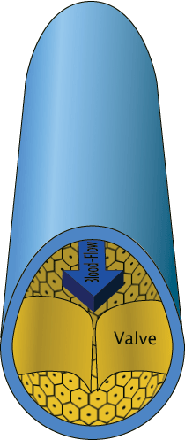
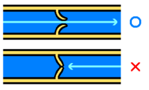
In anatomy, a vein is any of the blood vessels that carry blood toward the heart, most with one-way valves that prevent backflow. Veins are in contrast to the arteries, which are muscular blood vessels that carry blood away from the heart to the cells, tissues, and organs of the body. Most veins in the body carry deoxygenated blood from the tissues back to the heart, with the exception of the pulmonary and umbilical veins. The pulmonary vein carries oxygen-rich blood from the lungs to the left atrium of the heart, and the umbilical vein is present during fetal development and carries oxygenated blood from the placenta to the growing fetus.
The veins work in harmony with the arteries to produce a unified system for transporting blood with oxygen and nutrients to the cells, removing carbon dioxide and other cellular waste products, circulating hormones, lipoproteins, enzymes, and immune cells, and returning the blood to the heart.
The term "vein" has diverse meanings in other contexts. In botany, vein refers to the vascular tissue of leaves, located in the spongy layer of the mesophyll, that forms a branching framework of supporting and connecting tissue. The pattern of the veins is called venation. In zoology, veins are a supporting structure in an insect wing. In geology, a vein is a finite volume within a rock, having a distinct shape, filled with crystals of one or more minerals. This article will be limited to the use of the term with reference to the circulatory system.
Overview
The venal system is the lower-pressureâand normally lower oxygen-carryingâportion of the circulatory system. In the post-fetal human body, with the exception of the pulmonary vein, low oxygen blood moves from the capillaries of the arterial system to small, threadlike veins known as venules, which drain blood directly from the capillary beds, and from these the blood moves to larger and larger veins until back to the heart.
The arteries are perceived as carrying oxygenated blood to the tissues, while veins carry deoxygenated blood back to the heart. This is true of the systemic circulation, by far the larger of the two circuits of blood in the body, which transports oxygen from the heart to the tissues of the body. In pulmonary circulation, however, the arteries carry deoxygenated blood from the heart to the lungs and veins return oxygenated blood from the lungs to the heart. The difference between veins and arteries is their direction of flow (out of the heart by arteries, returning to the heart for veins), not their oxygen content. In addition, deoxygenated blood that is carried from the tissues back to the heart for reoxygenation in systemic circulation still carries some oxygen, though it is considerably less than that carried by the systemic arteries or pulmonary veins.
Anatomy
Like the arteries, the veins are defined by their three-layer walls, but the vein walls are less muscular and thinner than the artery walls. Skeletal muscle contractions help move the blood through the veins. The interiors of the larger veins are occupied by periodically occurring one-way flaps called venous valves, which prevent blood from flowing backward and from pooling in the lower extremities due to the effects of gravity. In humans, valves are absent in the smallest veins and most numerous in the extremities.
Excepting the pulmonary vein, veins function to return deoxygenated blood to the heart and are essentially tubes that collapse when their lumens are not filled with blood. The thick, outer-most layer of a vein is made of collagen, wrapped in bands of smooth muscle while the interior is lined with endothelial cells called intima. The precise location of veins is much more variable from person to person than that of arteries.
The total capacity of the veins in humans is more than sufficient to hold the entire blood volume of the body. This capacity is reduced through the venous tone of the smooth muscles, minimizing the cross-sectional area (and hence volume) of the individual veins and therefore total venous system. The helical bands of smooth muscles that wrap around veins help maintain blood flow to the right atrium. In cases of vasovagal syncope, the most common type of fainting, the smooth muscles relax and the veins of the extremities below the heart fill up with blood, failing to return sufficient volume to maintain cardiac output and blood flow to the brain.
Function
Veins return blood from organs to the heart. In systemic circulation in human beings, oxygenated blood is pumped by the left ventricle through the arteries to the muscles and organs of the body, where nutrients and oxygen in the blood are exchanged at capillaries for cellular wastes carbon dioxide. The deoxygenated and waste-laden blood flows through the veins to the right atrium of the heart, which transfers the blood to the right ventricle, from where it is pumped through the pulmonary arteries to the lungs. In pulmonary circulation the pulmonary veins return oxygenated blood from the lungs to the left atrium, which empties into the left ventricle, completing the cycle of blood circulation. (The cellular wastes are removed primarily by the kidneys.)
The return of blood to the heart is assisted by the action of the skeletal-muscle pump, which helps maintain the extremely low blood pressure of the venous system. Fainting can be caused by failure of the skeletal-muscular pump. Long periods of standing can result in blood pooling in the legs, with blood pressure too low to return blood to the heart. Neurogenic and hypovolaemic shock can also cause fainting. In these cases, the smooth muscles surrounding the veins become slack and the veins fill with the majority of the blood in the body, keeping blood away from the brain and causing unconsciousness.
In a functional analogy, the term "venous" in economics refers to recycling industries, in contrast to "arterial" or production industries.
Medical interest
Veins are used medically as points of access to the blood stream, permitting the withdrawal of blood specimens (venipuncture) for testing purposes, and intravenous delivery of fluid, electrolytes, nutrition, and medications through injection with a syringe, or by inserting a catheter. In contrast to arterial blood, which is uniform throughout the body, the blood removed from veins for testing can vary in its contents depending on the part of the body the vein drains; blood drained from a working muscle will contain significantly less oxygen and glucose than blood drained from the liver. However, the more blood from different veins mixes as it returns to the heart, the more homogeneous it becomes.
If an intravenous catheter has to be inserted, for most purposes this is done into a peripheral vein near the surface of the skin in the hand or arm, or less desirably, the leg. Some highly concentrated fluids or irritating medications must flow into the large central veins, which are sometimes used when peripheral access cannot be obtained. Catheters can be threaded into the superior vena cava for these uses: if long term use is thought to be needed, a more permanent access point can be inserted surgically.
Common diseases
The most common vein disorder is venous insufficiency, usually manifested by spider veins or varicose veins. A variety of treatments are used depending on the patient's particular type and pattern of veins and on the physician's preferences. Treatment can include radio-frequency ablation, vein stripping, ambulatory phlebectomy, foam sclerotherapy, lasers, or compression.
Deep vein thrombosis is a condition where a blood clot forms in a deep vein, which can lead to pulmonary embolism and chronic venous insufficiency.
Phlebology
Phlebology is the medical discipline that involves the diagnosis and treatment of disorders of venous origin. Diagnostic techniques used include the history and physical examination, venous imaging techniques, and laboratory evaluation related to venous thromboembolism. The American Medical Association has added phlebology to its list of Self-Designated Practice Specialties.
The American College of Phlebology is a professional organization of physicians and health care professionals from a variety of backgrounds. Annual meetings are conducted to facilitate learning and sharing of knowledge regarding venous disease. The equivalent body for countries in the Pacific is the Australasian College of Phlebology, active in Australia and New Zealand.
Notable veins and vein systems
The Great Saphenous vein (GSV) is the most important superficial vein of the lower limb of humans. First described by the Persian physician Avicenna, Saphenous derives its name from Safina, meaning hidden. This vein is "hidden" in its own fascial compartment in the thigh and only exits the fascia near the knee. Incompetence of this vein is an important cause of varicose veins of lower limbs.
The pulmonary veins carry relatively oxygenated blood from the lungs to the heart. The superior and inferior venae cavae carry relatively deoxygenated blood from the upper and lower systemic circulations, respectively.
A portal venous system is a series of veins or venules that directly connect two capillary beds. Examples of such systems include the hepatic portal vein and hypophyseal portal system.
Types of veins
Veins can be classified into:
- Portal vein vs. non-portal (most common)
- Superficial veins vs. deep veins
- Pulmonary veins vs. systemic veins
List of important named veins
- Jugular veins
- Pulmonary veins
- Portal vein
- Superior vena cava
- Inferior vena cava
- Iliac vein
- Femoral vein
- Popliteal vein
- Great saphenous vein
- Small saphenous vein
Names of important venule systems
- Portal venous system
- Systemic venous system
ReferencesISBN links support NWE through referral fees
- American College of Phlebology. n.d. What is phebology. American College of Phlebology. Retrieved May 3, 2008.
- Smith, P.C. 2004. Phlebology. Medi-data.co.uk. Retrieved May 3, 2008.
- Trupie, A.G.G. 2008. Veins: Introduction. Merck Manual. Retrieved May 3, 2008.
| ||||||||||||||||||
| ||||||||||||||
| |||||||||||||||||||||||
| ||||||||||||||
| Cardiovascular system - edit |
|---|
| Blood  | Heart â Aorta â Arteries â Arterioles â Capillaries â Venules â Veins â Vena cava â Heart â Pulmonary arteries â Lungs â Pulmonary veins â Heart |
Credits
New World Encyclopedia writers and editors rewrote and completed the Wikipedia article in accordance with New World Encyclopedia standards. This article abides by terms of the Creative Commons CC-by-sa 3.0 License (CC-by-sa), which may be used and disseminated with proper attribution. Credit is due under the terms of this license that can reference both the New World Encyclopedia contributors and the selfless volunteer contributors of the Wikimedia Foundation. To cite this article click here for a list of acceptable citing formats.The history of earlier contributions by wikipedians is accessible to researchers here:
The history of this article since it was imported to New World Encyclopedia:
Note: Some restrictions may apply to use of individual images which are separately licensed.

