Hindbrain
| Brain: Rhombencephalon | ||
|---|---|---|
 | ||
| Diagram depicting the main subdivisions of the embryonic vertebrate brain. These regions will later differentiate into forebrain, midbrain and hindbrain structures. | ||
 | ||
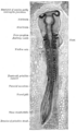
| Scheme of roof of fourth ventricle. | ||
| Gray's | subject #187 767 | |
| NeuroNames | hier-531 | |
| MeSH | Rhombencephalon | |
| Dorlands/Elsevier | r_12/12709581 | |
The rhombencephalon (or hindbrain) is a developmental categorization of portions of the central nervous system in vertebrates. It includes the medulla, pons, and cerebellum. Together they support vital bodily processes.[1]

The rhombencephalon can be subdivided in a variable number of transversal swellings called rhombomeres. In the human embryo eight rhombomeres can be distinguished, from caudal to rostral: Rh8-Rh1. Rostrally, the isthmus demarcates the boundary with the midbrain or mesencephalon.
A rare disease of the rhombencephalon, "rhombencephalosynapsis," is characterized by a missing vermis resulting in a fused cerebellum. Patients generally present with cerebellar ataxia.
The caudal rhombencephalon has been generally considered as the initiation site for neural tube closure.[2]
Myelencephalon
Rhombomeres Rh8-Rh4 form the myelencephalon.
The myelencephalon forms the medulla oblongata in the adult brain; it contains:
- a portion of the fourth ventricle,
- the glossopharyngeal nerve (CN IX),
- vagus nerve (CN X),
- accessory nerve (CN XI),
- hypoglossal nerve (CN XII),
- and a portion of the vestibulocochlear nerve (CN VIII).
Metencephalon
Rhombomeres Rh3-Rh1 form the metencephalon.
The metencephalon is composed of the pons and the cerebellum; it contains:
- a portion of the fourth ventricle,
- the trigeminal nerve (CN V),
- abducens nerve (CN VI),
- facial nerve (CN VII),
- and a portion of the vestibulocochlear nerve (CN VIII).
Evolution
The hindbrain is homologous to a part of the arthropod brain known as the sub-oesophageal ganglion, in terms of the genes that it expresses and its position in between the brain and the nerve cord.[3] On this basis, it has been suggested that the hindbrain first evolved in the Urbilaterian - the last common ancestor of chordates and arthropods - between 570 and 555 million years ago.[3][4]
Additional images
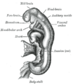
Chick embryo of thirty-three hours’ incubation, viewed from the dorsal aspect. X 30.
Embryo between eighteen and twenty-one days.
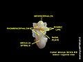
Rhombencephalon of human embryo


ReferencesISBN links support NWE through referral fees
- Haycock DE (2011). Being and Perceiving. Manupod Press. ISBN 978-0-9569621-0-2.
- ↑ Brain atlas - Hindbrain. Lundbeck Institute - Brain explorer. Retrieved 2013-06-04.
- ↑ SpringerLink - Journal Article
- ↑ 3.0 3.1 Ghysen A (2003). The origin and evolution of the nervous system. Int. J. Dev. Biol. 47 (7–8): 555–62.
- ↑ Haycock, DE Being and Perceiving
External links
- NIF Search - Rhombencephalon via the Neuroscience Information Framework
Template:Medulla Template:Pons Template:Fourth ventricle Template:Cerebellum
Credits
New World Encyclopedia writers and editors rewrote and completed the Wikipedia article in accordance with New World Encyclopedia standards. This article abides by terms of the Creative Commons CC-by-sa 3.0 License (CC-by-sa), which may be used and disseminated with proper attribution. Credit is due under the terms of this license that can reference both the New World Encyclopedia contributors and the selfless volunteer contributors of the Wikimedia Foundation. To cite this article click here for a list of acceptable citing formats.The history of earlier contributions by wikipedians is accessible to researchers here:
The history of this article since it was imported to New World Encyclopedia:
Note: Some restrictions may apply to use of individual images which are separately licensed.