Difference between revisions of "Hindbrain" - New World Encyclopedia
Rick Swarts (talk | contribs) |
Rick Swarts (talk | contribs) |
||
| Line 141: | Line 141: | ||
| url = http://books.google.co.uk/books?id=fXFeQb1z6bsC | | url = http://books.google.co.uk/books?id=fXFeQb1z6bsC | ||
}}</cite> | }}</cite> | ||
| + | |||
| + | Haycock DE (2011). Being and Perceiving. Manupod Press. ISBN 978-0-9569621-0-2. | ||
| + | |||
| + | <ref name=Ghysen>{{Cite journal|author=Ghysen A |title=The origin and evolution of the nervous system |journal=Int. J. Dev. Biol. |volume=47 |issue=7–8 |pages=555–62 |year=2003 |pmid=14756331 |doi= |url=http://www.ijdb.ehu.es/web/paper.php?doi=14756331}}</ref> | ||
| + | |||
<references/> | <references/> | ||
Revision as of 01:47, 9 December 2013
| Brain: Rhombencephalon | ||
|---|---|---|
 | ||
| Diagram depicting the main subdivisions of the embryonic vertebrate brain. These regions will later differentiate into forebrain, midbrain and hindbrain structures. | ||
 | ||
| Scheme of roof of fourth ventricle. | ||
| Gray's | subject #187 767 | |
| NeuroNames | hier-531 | |
| MeSH | Rhombencephalon | |
| Dorlands/Elsevier | r_12/12709581 | |
The rhombencephalon (or hindbrain) is a developmental categorization of portions of the central nervous system in vertebrates. It includes the medulla, pons, and cerebellum. Together they support vital bodily processes.[1]
The rhombencephalon can be subdivided in a variable number of transversal swellings called rhombomeres. In the human embryo eight rhombomeres can be distinguished, from caudal to rostral: Rh8-Rh1. Rostrally, the isthmus demarcates the boundary with the midbrain or mesencephalon.
A rare disease of the rhombencephalon, "rhombencephalosynapsis," is characterized by a missing vermis resulting in a fused cerebellum. Patients generally present with cerebellar ataxia.
The caudal rhombencephalon has been generally considered as the initiation site for neural tube closure.[2]
Overview

Vertebrate brains are characterized by three primary divisions: forebrain (or prosencephalon), midbrain (or mesencephalon), and hindbrain (or rhombencephalon).
The forebrain is dominant in terrestrial vertebrates, where it is the center of the processing sensor information. The forebrain of reptiles, amphibians, birds, and mammals is commonly divided into two regions: the "diencephalon," which consists of the hypothalamus and the thalamus, and the "telencephalon," or "end brain," which in mammals is called the cerebrum (Raven et al. 2008). The telencephalon also is the term used to refer to the embryonic structure from which the mature cerebrum develops.
The midbrain or mesencephalon is primarily is composed of the optic tectum, which processes and receives visual information.
The hindbrain or rhombencephalon includes the medulla oblongata, the pons, and the cerebellum. The hindbrain was the major component of early brains, as seen via casts of fossil agnathans, and remains the major part of fish brains today. The cerebellum carries on much of the coordination of motor reflexes (Raven et al. 2008), but is lacking in both hagfishes and lampreys (Northcutt 2002). The midbrain, pons, and medulla also are collectively referred to as the brainstem.
Developmentally, the hindbrain also can be subdivided into the myelencephalon, which is the area that gives way to development of the medulla oblongata, and the metencephalon, which gives rise to the pons and the cerebellum.
The hindbrain is homologous to a part of the arthropod brain known as the sub-oesophageal ganglion, in terms of the genes that it expresses and its position in between the brain and the nerve cord (Ghysen 2003). On this basis, it has been suggested that the hindbrain first evolved in the Urbilaterian—the last common ancestor of chordates and arthropods—between 570 and 555 million years ago (Ghysen 2003; Haycock 2011).
Myelencephalon: Medulla Oblongata

Rhombomeres Rh8-Rh4 form the myelencephalon.
The myelencephalon forms the medulla oblongata in the adult brain; it contains:
- a portion of the fourth ventricle,
- the glossopharyngeal nerve (CN IX),
- vagus nerve (CN X),
- accessory nerve (CN XI),
- hypoglossal nerve (CN XII),
- and a portion of the vestibulocochlear nerve (CN VIII).
The myelencephalon is a subdivision of the brain used to describe the area that gives way to development of the medulla oblongata. These are notes.
- The medulla, along with the spinal cord, contains many small nuclei involved in a wide variety of sensory and motor functions.[3]
Development
Myelencephalon to medulla oblongata
Order of brain development in fetus:
During fetal development, divisions that give rise to the hindbrain occur at just 28 days post conception with more specific subdivisions (metencephalon, myelencephalon) taking shape at 7 weeks post conception. Final shape differentiation into the medulla oblongata can be observed at 20 weeks gestation.[4]
Medulla oblongata
Function
The medulla oblongata serves as the connection to the spinal cord from the brain, together forming the Central Nervous System. The Central Nervous system is responsible for translating and responding to peripheral stimulus. The medulla regulates vasomotor function and autonomic responses such as respiration and cardiovascular systems as well as basic reflexive activities (coughing, sneezing, swallowing, vomiting).[5]
Damage/trauma
Because of its location at the brain stem, trauma to this area can be detrimental to survival of any kind. Research shows lesions resulting from trauma can cause pulmonary edemas due to the medullas association with pulmonary function.[6] Similarly, Ischemia can also result from lesions to the medulla affecting vasomotor function.[7]
Metencephalon: Pons and Cerebellum
Rhombomeres Rh3-Rh1 form the metencephalon.
The metencephalon is composed of the pons and the cerebellum; it contains:
- a portion of the fourth ventricle,
- the trigeminal nerve (CN V),
- abducens nerve (CN VI),
- facial nerve (CN VII),
- and a portion of the vestibulocochlear nerve (CN VIII).
Notes to be incorporated;
- The pons lies in the brainstem directly above the medulla. Among other things, it contains nuclei that control sleep, respiration, swallowing, bladder function, equilibrium, eye movement, facial expressions, and posture.[8]
- The cerebellum modulates the outputs of other brain systems to make them precise. Removal of the cerebellum does not prevent an animal from doing anything in particular, but it makes actions hesitant and clumsy. This precision is not built-in, but learned by trial and error. Learning how to ride a bicycle is an example of a type of neural plasticity that may take place largely within the cerebellum.[3]
The metencephalon is a developmental categorization of portions of the central nervous system. The metencephalon is composed of the pons and the cerebellum; contains a portion of the fourth ventricle; and the trigeminal nerve (CN V), abducens nerve (CN VI), facial nerve (CN VII), and a portion of the vestibulocochlear nerve (CN VIII).
Embryology
The metencephalon develops from the higher/rostral half of the embryonic rhombencephalon, and is differentiated from the myelencephalon in the embryo by approximately 5 weeks of age. By the third month, the metencephalon differentiates into its two main structures, the pons and the cerebellum.
Functions
The pons regulates breathing through particular nuclei that regulate the breathing center of the medulla oblongata. The cerebellum works to coordinate muscle movements, maintain posture, and integrate sensory information from the inner ear and proprioceptors in the muscles and joints.
Development
At the early stages of brain development, the brain vesicles that are formed are imperative.[9] Each brain region is characterized by its own specific architecture. These regions of the brain are determined by a combination of transcription factors and the signals that change their expression.[9]
The isthmus is the main organizing center for the tectum and the cerebellum.[10] The tectum is the dorsal part of the metencephalon. The tectum includes the superior and inferior colliculli which play a part in visual and audio processing. Two of the major genes that affect the metencephalon are Fgf8 and Wnt1, which are both expressed around the isthmus. Fgf8 is also known as Fibroblast Growth Factor 8. It is a protein that is widely thought to be the most important organizing signal. Its main function is to set up and maintain the barrier between the midbrain and hindbrain, specifically between the mesencephalon and metencephalon.[10] It also plays a large role in deciding the structure of the mid- and hindbrain. Wnt1 is a proto-oncogene protein (Wingless-type MMTV integration site family, member 1). This gene was originally thought to play a role in the development of the midbrain and hindbrain, but studies have shown that this may not be the case.[10] Wnt1 is thought to be behind the genetic disorder called Joubert Syndrome, a disorder that affects the cerebellum.
Otx1 and Otx2 are genes that play important parts in the development of the brain and studies have shown that their roles change throughout the brain’s development.[11] It is thought that at the stage of brain development where the rostral brain is regionalized into its different parts (telencephalon, diencephalon, metencephalon, and mesencephalon) that Otx2 and Otx1 protect the caudalization of the diencephalon and mesencephalon into metencephalon.[11]
Here is a list of some of the most important vertebrate brain components, along with a brief description of their functions as currently understood:
Additional images
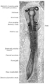
Chick embryo of thirty-three hours’ incubation, viewed from the dorsal aspect. X 30.
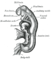
Embryo between eighteen and twenty-one days.
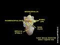
Rhombencephalon of human embryo


ReferencesISBN links support NWE through referral fees
- Haycock DE (2011). Being and Perceiving. Manupod Press. ISBN 978-0-9569621-0-2.
Haycock DE (2011). Being and Perceiving. Manupod Press. ISBN 978-0-9569621-0-2.
- ↑ Brain atlas - Hindbrain. Lundbeck Institute - Brain explorer. Retrieved 2013-06-04.
- ↑ SpringerLink - Journal Article
- ↑ 3.0 3.1 Cite error: Invalid
<ref>tag; no text was provided for refs namedKandel 2000 - ↑ Carlson, Neil R. Foundations of Behavioral Neuroscience.63-65
- ↑ Loewy, A. D., & Spyer, K. M. (Eds.). (1990). Central regulation of autonomic functions. Oxford University Press, USA.145-164
- ↑ Matsuyama, T., Okuchi, K., Nishiguchi, T., Seki, T., & Murao, Y. (2007). Neurogenic pulmonary edema caused by a medulla oblongata lesion after head trauma. The Journal of trauma, 63(3), 700.
- ↑ Kumada, M. A. M. O. R. U., Dampney, R. A., & Reis, D. J. (1979). Profound hypotension and abolition of the vasomotor component of the cerebral ischemic response produced by restricted lesions of medulla oblongata in rabbit. Relationship to the so-called tonic vasomotor center. Circulation research, 45(1), 63-70.
- ↑ Siegel, A (2010). Essential Neuroscience. Lippincott Williams & Wilkins, 184–189. ISBN 978-0-7817-8383-5.
- ↑ 9.0 9.1 Nakamura, H., and Watanabe, Y. Isthmus organizer and regionalization of the mesencephalon and metencephalon. Int. J. Dev. Biol. 49: 231-235 (2005). doi: 10.1387/ijdb.041964hn.
- ↑ 10.0 10.1 10.2 Matsunaga, E., Katahira, T., and Nakamura, H. "Role of Lmx1b and Wnt1 in mesencephalon and metencephalon development""Development" (2002)
- ↑ 11.0 11.1 Sakuri, Y., Kurokawa, D., Kiyonari, H., Kajikawa, E., Suda, Y., and Aizawa, S. "Otx2 and Otx1 protect diencephalon and mesencephalon from caudalization into metencephalon during early brain regionalization" "Developmental Biology" (2010).
- ↑ Ghysen A (2003). The origin and evolution of the nervous system. Int. J. Dev. Biol. 47 (7–8): 555–62.
References
- Brink, T. L. 2008. Unit 4: The nervous system. In T. L. Brink, Psychology: A Student Friendly Approach. Retrieved November 16, 2013.
- Campbell, N. A., J. B. Reece, L. A. Urry, et al. 2008. Biology, 8th edition. San Francisco: Pearson/Benjamin Cummings. ISBN 9780805368444.
- Chamberlin, S. L., and B. Narins. 2005. The Gale Encyclopedia of Neurological Disorders. Detroit: Thomson Gale. ISBN 078769150X.
- Dafny, N. 2013. chapter 1: Overview of the Nervous system. Neuroscience Online. Retrieved November 15, 2013.
- Freudenrich, C., and R. Boyd. 2013. How your brain works. How Stuff Works. Retrieved November 16, 2013.
- Gonzalez, R. 2012. The 4 biggest myths about the human brain. 109.com. Retrieved November 12, 2013.
- Marieb, E. N. and K. Hoehn. 2010. Human Anatomy & Physiology, 8th edition. Benjamin Cummings. ISBN 9780805395693.
- Northcutt, R. G. 2002. Understanding vertebrate brain evolution. Integ. and Comp. Biol. 42: 743-756.
- Raven, P. H., G. B. Johnson, J. B. Losos, K. A. Mason, and S. R. Singer. 2008. Biology, 8th edition. Boston: McGraw Hill. ISBN 9780072965810.
- Ruppert, E. E., R. S. Fox, and R. D. Barnes. 2004. Invertebrate Zoology, 7 ed. Brooks/Cole. ISBN 0030259827.
External links
- NIF Search - Rhombencephalon via the Neuroscience Information Framework
Template:Medulla Template:Pons Template:Fourth ventricle Template:Cerebellum
Credits
New World Encyclopedia writers and editors rewrote and completed the Wikipedia article in accordance with New World Encyclopedia standards. This article abides by terms of the Creative Commons CC-by-sa 3.0 License (CC-by-sa), which may be used and disseminated with proper attribution. Credit is due under the terms of this license that can reference both the New World Encyclopedia contributors and the selfless volunteer contributors of the Wikimedia Foundation. To cite this article click here for a list of acceptable citing formats.The history of earlier contributions by wikipedians is accessible to researchers here:
The history of this article since it was imported to New World Encyclopedia:
Note: Some restrictions may apply to use of individual images which are separately licensed.