Sympathetic nervous system
| Brain: Sympathetic nervous system | ||
|---|---|---|
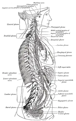
| The sympathetic nervous system extends from the thoracic to lumbar vertebrae and has connections with the thoracic,cat, and pelvic plexuses. | ||
| Latin | pars sympathica divisionis autonomici systematis nervosi | |
The sympathetic nervous system (SNS) is one of the main subdivisions of the autonomic nervous system (ANS) of vertebrates; often known as the "fight or flight system," it functions in energy generation and arousal, helping to mobilize the body during times of excitement, stress, and when activity and a quick response might be needed. For example, during threatening situations, the SNS can accelerate the heart, dilate the eyes' pupils, constrict visceral blood vessels, shunt blood to active skeletal muscles, inhibit activity of the stomach and intestine, dilate the bronchioles in the lung, inhibit the emptying of the bladder, and release glucose from the liver. Its general action is often in contrast with the ANS' other main subdivision , the parasympathetic nervous system (PSNS), which provides a more calming, energy conserving, self-maintenance, "rest and digest" function during time of rest, such as lowering metabolic rate, synthesizing glycogen, and restoring blood pressure and resting heartbeat. A third subsystem of the ANS is the enteric nervous system.
The sympathetic and parasympathetic systems create balance for the body via offering opposite and yet complementary effects, each essential to respond to appropriate conditions. In response to stress and danger, the sympathetic nervous system releases epinephrines (adrenaline), and in general increases conditions for activity and higher metabolic rate. During times of rest, sleeping, and digesting food, the parasympathetic nervous system counters this, releasing acetylcholine, and in general slows activity, lowers metabolic rate, restores blood pressure, enhances digestion, and so forth.
However, only in extreme cases, such as being threatened versus sleeping, would one visualize one ANS branch or the other dominating so strongly. In reality, the SNS and PSNS work in tandem to create a synergistic stimulation that is not merely on or off, but can be described as a continuum depending upon how vigorously each division is attempting to carry out its actions. And in the case of human reproductive activity, the two divisions strongly complement the actions of each other. In a sense, the actions of the SNS and PSNS is reflective of the complementary, interdependent forces described by the ancient Chinese philosophy yin and yang, whereby both forces are necessary to create overall harmony and balance in the living organism.
Overview of vertebrate nervous system
The sympathetic nervous system is a main subsystem of the autonomic nervous system (ANS), which in turn is part of the peripheral nervous system (PNS).
The vertebrate nervous system is divided into the central nervous system (CNS), comprising the brain and spinal cord, and the peripheral nervous system (PNS), consisting of all the nerves and neurons that reside or extend outside the central nervous system, such as to serve the limbs and organs.
The peripheral nervous system, in turn, is commonly divided into two subsystems, the somatic nervous system and the autonomic nervous system. The somatic nervous system (or sensory-somatic nervous system) involves nerves just under the skin and serves as the sensory connection between the outside environment and the CNS. These nerves are under conscious control, but most have an automatic component, as is seen in the fact that they function even in the case of a coma (Anissimov 2007). In humans, the somatic nervous system consists of 12 pairs of cranial nerves and 31 pairs of spinal nerves (Chamberlin and Narins 2005).
The autonomic nervous system is typically presented as that portion of the peripheral nervous system that is independent of conscious control, acting involuntarily and subconsciously (reflexively), and innervating heart muscle, endocrine glands, exocrine glands, and smooth muscle (Chamberlin and Narins 2005). In contrast, the somatic nervous system innervates skeletal muscle tissue, rather than smooth, cardiac, or glandular tissue.
The autonomic nervous system is subdivided into the sympathetic nervous system, the parasympathetic nervous system, and the enteric nervous system. In general, the sympathetic nervous system increases activity and metabolic rate (the "fight or flight response"), while the parasympathetic slows activity and metabolic rate, returning the body to normal levels of function (the "rest and digest state") after heightened activity from sympathetic stimulation (Chamberlin and Narins 2005). The enteric nervous system innervates areas around the intestines, pancreas, and gall bladder, dealing with digestion, and so forth.
Unlike the somatic nervous system, which always excites muscle tissue, the autonomic nervous system can either excite or inhibit innervated tissue (Chamberlin and Narins 2005). Most associated tissues and organs have nerves of both the sympathetic and the parasympathetic nervous systems. The two system can stimulate the target organs and tissues in opposite ways, such as sympathetic stimulation to increase heart rate and parasympathetic to decrease heart rate, or the sympathetic stimulation resulting in pupil dilation, and the parasympathetic in pupil constriction or narrowing (Chamberlin and Narins 2005). Or, they can both stimulate activity in concert, but in different ways, such as both increasing saliva production by salivary glands, but with sympathetic stimulation yielding viscous or thick saliva and parasympathetic yielding watery saliva.
The autonomic nervous system is that part of the peripheral nervous system that largely acts independent of conscious control (involuntarily) and consists of nerves in cardiac muscle, smooth muscle, and exocrine and endocrine glands. The other main subdivision of the peripheral nervous system, the somatic nervous system, consists of cranial and spinal nerves that innervate skeletal muscle tissue, rather than smooth, cardiac, or glandular tissue, and this portion is considered to be more under voluntary control (Anissimov 2006; Towle 1989).
In addition to the parasympathetic nervous system, the other main subdivision of the autonomic nervous system is the sympathetic nervous system. The enteric nervous system commonly also is considered a subdivision of the autonomic nervous system.
In sending fibers to three tissues—cardiac muscle, smooth muscle, or glandular tissue—the autonomic nervous system provides stimulation, sympathetic or parasympathetic, to control smooth muscle contraction, regulate cardiac muscle, or stimulate or inhibit glandular secretion.
While sympathetic nervous system and parasympathetic divisions typically function in opposition to each other, this opposition is better understood as complementary in nature rather than antagonistic. For an analogy, one may think of the sympathetic division as the accelerator and the parasympathetic division as the brake. The sympathetic division typically functions in actions requiring quick responses. The parasympathetic division functions with actions that do not require immediate reaction. A rarely used (but useful) acronym used to summarize the functions of the parasympathetic nervous system in human beings is SLUDD (salivation, lacrimation, urination, digestion, and defecation).
E division (excerise, excitement, emegey , embarrassment
D division (digestion, defecation, diuresis (urination)
Overview
Alongside the two components of the autonomic nervous system, the sympathetic nervous system aids in the control of most of the body's internal organs. Stress—as in the flight-or-fight response—is thought to counteract the parasympathetic system, which generally works to promote maintenance of the body at rest. In truth, the functions of both the parasympathetic and sympathetic nervous systems are not so straightforward, but this is a useful rule of thumb.[1][2]
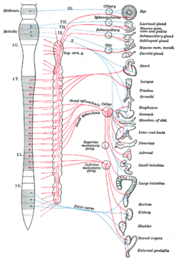
There are two kinds of neurons involved in the transmission of any signal through the sympathetic system: pre- and post-ganglionic. The shorter preganglionic neurons originate from the thoracolumbar region of the spinal cord (levels T1 - L3, specifically) and travel to a ganglion, often one of the paravertebral ganglia, where they synapse with a postganglionic neuron. From there, the long postganglionic neurons extend across most of the body.[3]
At the synapses within the ganglia, preganglionic neurons release acetylcholine, a neurotransmitter that activates nicotinic acetylcholine receptors on postganglionic neurons. In response to this stimulus postganglionic neurons - with two important exceptions - release norepinephrine, which activates adrenergic receptors on the peripheral target tissues. The activation of target tissue receptors causes the effects associated with the sympathetic system.[4]
The two exceptions mentioned above are postganglionic neurons of sweat glands and chromaffin cells of the adrenal medulla. Postganglionic neurons of sweat glands release acetylcholine for the activation of muscarinic receptors. Chromaffin cells of the adrenal medulla are analogous to post-ganglionic neurons; the adrenal medulla develops in tandem with the sympathetic nervous system and acts as a modified sympathetic ganglion. Within this endocrine gland, pre-ganglionic neurons synapse with chromaffin cells, stimulating the chromaffin to release norepinephrine and epinephrine directly into the blood.[5]
Function
| Organ | Effect |
|---|---|
| Eye | Dilates pupil |
| Heart | Increases rate and force of contraction |
| Lungs | Dilates bronchioles |
| Blood Vessels | Constricts |
| Sweat Glands | Activates sweat secretion |
| Digestive tract | Inhibits peristalsis |
| Kidney | Increases renin secretion |
| Penis | Promotes ejaculation |
The sympathetic nervous system is responsible for up- and down-regulating many homeostatic mechanisms in living organisms. Fibers from the SNS innervate tissues in almost every organ system, providing at least some regulatory function to things as diverse as pupil diameter, gut motility, and urinary output. It is perhaps best known for mediating the neuronal and hormonal stress response commonly known as the fight-or-flight response. This response is also known as sympatho-adrenal response of the body, as the preganglionic sympathetic fibers that end in the adrenal medulla (but also all other sympathetic fibers) secrete acetylcholine, which activates the great secretion of adrenaline (epinephrine) and to a lesser extent noradrenaline (norepinephrine) from it. Therefore, this response that acts primarily on the cardiovascular system is mediated directly via impulses transmitted through the sympathetic nervous system and indirectly via catecholamines secreted from the adrenal medulla.
Some evolutionary theorists suggest that the sympathetic nervous system operated in early organisms to maintain survival as the sympathetic nervous system is responsible for priming the body for action.[6] One example of this priming is in the moments before waking, in which sympathetic outflow spontaneously increases in preparation for action.
Organization
Sympathetic nerves originate inside the vertebral column, toward the middle of the spinal cord in the intermediolateral cell column (or lateral horn), beginning at the first thoracic segment of the spinal cord and are thought to extend to the second or third lumbar segments. Because its cells begin in the thoracic and lumbar regions of the spinal cord, the SNS is said to have a thoracolumbar outflow. Axons of these nerves leave the spinal cord through the anterior rootlet/root. They pass near the spinal (sensory) ganglion, where they enter the anterior rami of the spinal nerves. However, unlike somatic innervation, they quickly separate out through white rami connectors (so called from the shiny white sheaths of myelin around each axon) that connect to either the paravertebral (which lie near the vertebral column) or prevertebral (which lie near the aortic bifurcation) ganglia extending alongside the spinal column.
To reach target organs and glands, the axons must travel long distances in the body, and, to accomplish this, many axons relay their message to a second cell through synaptic transmission. The ends of the axons link across a space, the synapse, to the dendrites of the second cell. The first cell (the presynaptic cell) sends a neurotransmitter across the synaptic cleft where it activates the second cell (the postsynaptic cell). The message is then carried to the final destination.
Presynaptic nerves' axons terminate in either the paravertebral ganglia or prevertebral ganglia. There are four different ways an axon can take before reaching its terminal. In all cases, the axon enters the paravertebral ganglion at the level of its originating spinal nerve. After this, it can then either synapse in this ganglion, ascend to a more superior or descend to a more inferior paravertebral ganglion and synapse there, or it can descend to a prevertebral ganglion and synapse there with the postsynaptic cell.
The postsynaptic cell then goes on to innervate the targeted end effector (i.e. gland, smooth muscle, etc.). Because paravertebral and prevertebral ganglia are relatively close to the spinal cord, presynaptic neurons are generally much shorter than their postsynaptic counterparts, which must extend throughout the body to reach their destinations.
A notable exception to the routes mentioned above is the sympathetic innervation of the suprarenal (adrenal) medulla. In this case, presynaptic neurons pass through paraverterbral ganglia, on through prevertebral ganglia and then synapse directly with suprarenal tissue. This tissue consists of cells that have pseudo-neuron like qualities in that when activated by the presynaptic neuron, they will release their neurotransmitter (epinephrine) directly into the blood stream.
In the SNS and other components of the peripheral nervous system, these synapses are made at sites called ganglia. The cell that sends its fiber is called a preganglionic cell, while the cell whose fiber leaves the ganglion is called a postganglionic cell. As mentioned previously, the preganglionic cells of the SNS are located between the first thoracic segment and third lumbar segments of the spinal cord. Postganglionic cells have their cell bodies in the ganglia and send their axons to target organs or glands.
The ganglia include not just the sympathetic trunks but also the cervical ganglia (superior, middle and inferior), which sends sympathetic nerve fibers to the head and thorax organs, and the celiac and mesenteric ganglia (which send sympathetic fibers to the gut).
Sensation
The afferent fibers of the autonomic nervous system, which transmit sensory information from the internal organs of the body back to the central nervous system, are not divided into parasympathetic and sympathetic fibers as the efferent fibers are.[7] Instead, autonomic sensory information is conducted by general visceral afferent fibers.
General visceral afferent sensations are mostly unconscious visceral motor reflex sensations from hollow organs and glands that are transmitted to the CNS. While the unconscious reflex arcs normally are undetectable, in certain instances they may send pain sensations to the CNS masked as referred pain. If the peritoneal cavity becomes inflamed or if the bowel is suddenly distended, the body will interpret the afferent pain stimulus as somatic in origin. This pain is usually non-localized. The pain is also usually referred to dermatomes that are at the same spinal nerve level as the visceral afferent synapse.[citation needed]
Information transmission
Messages travel through the SNS in a bidirectional flow. Efferent messages can trigger changes in different parts of the body simultaneously. For example, the sympathetic nervous system can accelerate heart rate; widen bronchial passages; decrease motility (movement) of the large intestine; constrict blood vessels; increase peristalsis in the esophagus; cause pupillary dilation, piloerection (goose bumps) and perspiration (sweating); and raise blood pressure.
The first synapse (preganglionic neuron to postganglionic neuron in the sympathetic chain) is mediated by nicotinic receptors physiologically activated by acetylcholine. The target synapse of the postganglionic neuron is mediated by adrenergic receptors and is physiologically activated by either noradrenaline (norepinephrine) or adrenaline (epinephrine). An exception is with sweat glands, which receive sympathetic innervation but have muscarinic acetylcholine receptors, which are normally characteristic of Parasympathetic nervous system. Another exception is with certain deep muscle blood vessels, which dilate (rather than constrict) with an increase in sympathetic tone. This is because of the presence of more beta2 receptors (rather than alpha1, which are frequently found on other vessels)As it inhibits peristalsis it is clear that it concentrates blood flow towards periphery of body.
Sympathicotonia
Sympathicotonia is a stimulated[8] condition of the sympathetic nervous system, marked by vascular spasm,[9] elevated blood pressure,[9] and goose bumps.[9]
See also
- Epinephrine
- History of catecholamine research
- Norepinephrine
- Sympathetic ganglia
- Sympathetic trunk
ReferencesISBN links support NWE through referral fees
- ↑ Cite error: Invalid
<ref>tag; no text was provided for refs namedbrodal - ↑ Sherwood, Lauralee (2008). Human Physiology: From Cells to Systems, 7, Cengage Learning, 240. ISBN 0-495-39184-0.
- ↑ (2005) Gray's Anatomy for Students, 1, Elsevier, 76–84. ISBN 0-443-06612-4.
- ↑ (2007) Rang and Dale's Pharmacology, 6, Elsevier, 135. ISBN 0-443-06911-5.
- ↑ 5.0 5.1 Silverthorn, Dee Unglaub (2009). Human Physiology: An Integrated Approach, 4, Pearson/Benjamin Cummings, 379–386. ISBN 0-321-54130-8.
- ↑ Robert Ornstein (1992). The Evolution of Consciousness: of Darwin, Freud, and Cranial Fire: The Origins of the Way We Think. New York: Simon & Schuster. ISBN 0-671-79224-5.
- ↑ Moore, K.L., & Agur, A.M. (2007). Essential Clinical Anatomy: Third Edition. Baltimore: Lippincott Williams & Wilkins. 34-35. ISBN 978-0-7817-6274-8
- ↑ thefreedictionary.com Citing: Dorland's Medical Dictionary for Health Consumers. © 2007
- ↑ 9.0 9.1 9.2 thefreedictionary.com Citing: The American Heritage Medical Dictionary Copyright © 2007
Template:Autonomic
Sympathetic_nervous_system&oldid=560024815
Credits
New World Encyclopedia writers and editors rewrote and completed the Wikipedia article in accordance with New World Encyclopedia standards. This article abides by terms of the Creative Commons CC-by-sa 3.0 License (CC-by-sa), which may be used and disseminated with proper attribution. Credit is due under the terms of this license that can reference both the New World Encyclopedia contributors and the selfless volunteer contributors of the Wikimedia Foundation. To cite this article click here for a list of acceptable citing formats.The history of earlier contributions by wikipedians is accessible to researchers here:
The history of this article since it was imported to New World Encyclopedia:
Note: Some restrictions may apply to use of individual images which are separately licensed.

