Peripheral nervous system
The peripheral nervous system (PNS) is that portion of the vertebrate nervous system that is outside the brain and spinal cord, which comprise the central nervous system (CNS). The peripheral nervous system connects with the central nervous system, serving as a conduit for neural signals to be transmitted to and from the CNS to the body. Unlike the CNS, the PNS is not protected by bone or the blood-brain barrier, leaving it exposed to toxins and mechanical injuries.
The peripheral nervous system, in combination with the central nervous system, allows humans beings to harmonize with their internal and external environments. Without the peripheral nervous system, one could not sense the external environment (smell, sight, touch, taste, hear), and would neither recognize threats or pleasurable experiences. One would not even know if it were light or dark, rainy or sunny, hot or cold. Internally, human organs and organ systems would not be able to coordinate, but would act independently; muscle movement and glandular activity would be chaotic.
The peripheral nervous system is divided into two subsystems, the somatic nervous system and the autonomic nervous system. The autonomic nervous system is further subdivided into the sympathetic nervous system, the parasympathetic nervous system, and the enteric nervous system.
Overview
The nervous system is that network of specialized cells, tissues, and organs that coordinates the body's interaction with the environment, such as sensing the environment, monitoring organs, and coordinating the activity of muscles. The nervous system of vertebrate animals is divided into the central nervous system and the peripheral nervous system (PNS). The CNS comprises the brain and spinal cord, whereas the PNS consists of the nerves and neurons that reside or extend outside the central nervous system, such as to serve the limbs and organs.
| Neuron |
|---|
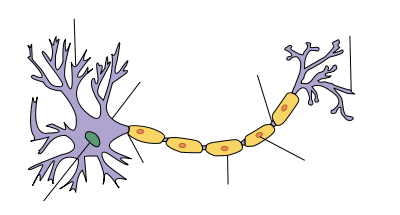
| Structure of a typical neuron |
All parts of the nervous system are made of nervous tissue, which conducts electrical impulses. Prominent components in a nervous system include neurons (nerve cells) and nerves. Neurons are typically composed of a soma, or cell body, a dendritic tree, and an axon. The large majority of what are commonly called nerves (which are actually bundles of axonal processes of nerve cells) are considered to be PNS.
Neurons active in the PNS can be divided into sensory neurons and motor neurons (Chamberlin and Narins 2005). Sensory neurons act as a conduit between sensory receptors that sense stimuli such as cold, heat, and pain and the CNS. Motor neurons act as a conduit between the CNS and various muscles and glands.
The peripheral nervous system is commonly subdivided into two subsystems, the somatic nervous system and the autonomic nervous system.
The somatic nervous system involves nerves just under the skin and serves as the connection between the outside environment and the CNS. These nerves are under conscious control, but there still is a largely automatic component. For example, they function even in the case of a coma (Anissimov 2007). In humans, the somatic nervous system consists of 12 pairs of cranial nerves and 31 pairs of spinal nerves (Chamberlin and Narins 2005). Some pairs are exclusively sensory, such as those used for smell, vision, hearing, and balance; some are strictly motor neurons, such as for movement of eyeballs, swallowing, and movement of the head; and some have sensory and motor neurons working in tandem, such as with the sense of taste. The spinal neurons all contain both motor and sensory neurons (Chamberlin and Narins 2005).
The autonomic nervous system is subdivided into the sympathetic nervous system, the parasympathetic nervous system, and the enteric nervous system. The autonomic nervous systems have a reputation as independent of conscious control, acting involuntarily (reflexively), responsible for functions of the heart muscle, endocrine glands, exocrine glands, and smooth muscle (Chamberin and Narins 2005). The sympathetic nervous system deals with the response to stress and danger, the release of epinephrine (adrenaline), and in general increases activity and metabolic rate, innervating cardiac muscle, muscle, and glandular tissue. The parasympathetic nervous system is central during rest, sleeping, and digestion, and in general lowers metabolic rate and slows activity, innervating the same types of tissues as the sympathetic nerves but restoring blood pressure, resting heartbeat, and so forth (Chamberlain and Narins 2005). The enteric nervous system has nerves around the intestines, pancreas, and gallbladder.
The hypothalamus controls the involuntary autonomic nervous system, while other regions of the brain regulates the somatic nervous system.
Naming of specific nerves
Ten out of the twelve cranial nerves originate from the brainstem, and mainly control the functions of the anatomic structures of the head with some exceptions. CN X (10) receives visceral sensory information from the thorax and abdomen, and CN XI (11) is responsible for innervating the sternocleidomastoid and trapezius muscles, neither of which is exclusively in the head.
Spinal nerves take their origins from the spinal cord. They control the functions of the rest of the body. In humans, there are 31 pairs of spinal nerves: 8 cervical, 12 thoracic, 5 lumber, 5 sacral, and 1 coccygeal. The naming convention for spinal nerves is to name it after the vertebra immediately above it. Thus, the fourth thoracic nerve originates just below the fourth thoracic vertebra. This convention breaks down in the cervical spine. The first spinal nerve originates above the first cervical vertebra and is called C1. This continues down to the last cervical spinal nerve, C8. There are only 7 cervical vertebrae and 8 cervical spinal nerves.
Cervical spinal nerves (C1-C4)
The first 4 cervical spinal nerves, C1 through C4, split and recombine to produce a variety of nerves that subserve the neck and back of head.
Spinal nerve C1 is called the suboccipital nerve, which provides motor innervation to muscles at the base of the skull.
C2 and C3 form many of the nerves of the neck, providing both sensory and motor control. These include the greater occipital nerve, which provides sensation to the back of the head, the lesser occipital nerve, which provides sensation to the area behind the ears, the greater auricular nerve and the lesser auricular nerve.
The phrenic nerve arises from nerve roots C3, C4, and C5. It innervates the diaphragm, enabling breathing. If the spinal cord is transected above C3, then spontaneous breathing is not possible.
Brachial plexus (C5-T1)
The last 4 cervical spinal nerves, C5 through C8, and the first thoracic spinal nerve, T1, combine to form the brachial plexus, or plexus brachialis, a tangled array of nerves, splitting, combining, and recombining to form the nerves that subserve the arm and upper back. Although the brachial plexus may appear tangled, it is highly organized and predictable, with little variation between people.
The first nerve off the brachial plexus is the dorsal scapular nerve, arising from C5 nerve root and innervating the rhomboids and the levator scapulae muscles. The long thoracic nerve arises from C5, C6, and C7 to innervate the serratus anterior.
The brachial plexus first forms three trunks, the superior trunk, composed of the C5 and C6 nerve roots, the middle trunk, made of the C7 nerve root, and the inferior trunk, made of the C8 and T1 nerve roots. The suprascapular nerve is an early branch of the superior trunk. It innervates the suprascapular and infrascapular muscles, part of the rotator cuff.
The trunks reshuffle as they traverse towards the arm into cords. There are three of them. The lateral cord is made up of fibers from the superior and middle trunk. The posterior cord is made up of fibers from all three trunks. The medial cord is composed of fibers solely from the medial trunk.
Lateral cord
The lateral cord gives rise to the following nerves:
- The lateral pectoral nerve, C5, C6, and C7 to the pectoralis major muscle, or musculus pectoralis major.
- The musculocutaneous nerve, which innervates the biceps muscle.
- The median nerve, partly. The other part comes from the medial cord.
Posterior cord
The posterior cord gives rise to the following nerves:
- The upper subscapular nerve, C7 and C8, to the subscapularis muscle, or musculus supca of the rotator cuff.
- The lower subscapular nerve, C5 and C6, to the teres major muscle, or the musculus teres major.
- The thoracodorsal nerve, C6, C7, and C8, to the latissimus dorsi muscle, or musculus latissimus dorsi.
- The axillary nerve, which supplies sensation to the shoulder and motor to the deltoid muscle or musculus deltoideus, and the teres minor muscle, or musculus teres minor, also of the rotator cuff.
- The radial nerve, or nervus radialis, which innervates the triceps brachii muscle, the brachioradialis muscle, or musculus brachioradialis, the extensor muscles of the fingers and wrist (extensor carpi radialis muscle), and the extensor and abductor muscles of the thumb.
Medial cord
The medial cord gives rise to the following nerves:
- The median pectoral nerve, C8 and T1, to the pectoralis muscle
- The medial brachial cutaneous nerve, T1
- The medial antebrachial cutaneous nerve, C8 and T1
- The median nerve, partly. The other part comes from the lateral cord, C7, C8, and T1 nerve roots. The first branch of the median nerve is to the pronator teres muscle, then the flexor carpi radialis, the palmaris longus and the flexor digitorum superficialis. The median nerve provides sensation to the anterior palm, the anterior thumb, index finger, and middle finger. It is the nerve compressed in carpal tunnel syndrome.
- The ulnar nerve originates in nerve roots C7, C8, and T1. It provides sensation to the ring and pinky fingers. It innervates the flexor carpi ulnaris muscle, the flexor digitorum profundus muscle to the ring and pinky fingers, and the intrinsic muscles of the hand (the interosseous muscle, the lumbrical muscles and the flexor pollicus brevis muscle). This nerve traverses a groove on the elbow called the cubital tunnel, also known as the funny bone. Striking the nerve at this point produces an unpleasant sensation in the ring and little fingers.
ReferencesISBN links support NWE through referral fees
- Anissimov, M. 2007. How does the nervous system work? Conjecture Corporation: Wise Geek. Retrieved May 13, 2007.
- Chamberlin, S. L., and B. Narins. 2005. The Gale Encyclopedia of Neurological Disorders. Detroit: Thomson Gale. ISBN 078769150X
- Towle, A. 1989. Modern Biology. Austin, TX: Holt, Rinehart and Winston. ISBN 0030139198
Credits
New World Encyclopedia writers and editors rewrote and completed the Wikipedia article in accordance with New World Encyclopedia standards. This article abides by terms of the Creative Commons CC-by-sa 3.0 License (CC-by-sa), which may be used and disseminated with proper attribution. Credit is due under the terms of this license that can reference both the New World Encyclopedia contributors and the selfless volunteer contributors of the Wikimedia Foundation. To cite this article click here for a list of acceptable citing formats.The history of earlier contributions by wikipedians is accessible to researchers here:
The history of this article since it was imported to New World Encyclopedia:
Note: Some restrictions may apply to use of individual images which are separately licensed.
