Difference between revisions of "Cardiac muscle" - New World Encyclopedia
Rick Swarts (talk | contribs) |
Rick Swarts (talk | contribs) |
||
| Line 48: | Line 48: | ||
| − | |||
| − | |||
'''Cardiac muscle''' is adapted to be highly resistant to fatigue: it has a large number of [[Mitochondrion|mitochondria]], enabling continuous [[aerobic respiration]], numerous [[myoglobin]]s ([[oxygen]]-storing pigment), and a good blood supply, which provides nutrients and oxygen. The heart is so tuned to aerobic metabolism that it is unable to pump sufficiently in [[ischaemia|ischaemic]] conditions. At [[basal metabolic rate]]s, about 1% of energy is derived from [[anaerobic metabolism]]. This can increase to 10% under moderately [[Hypoxia (medical)|hypoxic]] conditions, but, under more severe hypoxic conditions, not enough energy can be liberated by [[Lactic acid#Exercise and lactate|lactate production]] to sustain [[Ventricle (heart)|ventricular]] contractions.<ref>Ganong, Review of Medical Physiology, 22nd Edition. p81</ref> | '''Cardiac muscle''' is adapted to be highly resistant to fatigue: it has a large number of [[Mitochondrion|mitochondria]], enabling continuous [[aerobic respiration]], numerous [[myoglobin]]s ([[oxygen]]-storing pigment), and a good blood supply, which provides nutrients and oxygen. The heart is so tuned to aerobic metabolism that it is unable to pump sufficiently in [[ischaemia|ischaemic]] conditions. At [[basal metabolic rate]]s, about 1% of energy is derived from [[anaerobic metabolism]]. This can increase to 10% under moderately [[Hypoxia (medical)|hypoxic]] conditions, but, under more severe hypoxic conditions, not enough energy can be liberated by [[Lactic acid#Exercise and lactate|lactate production]] to sustain [[Ventricle (heart)|ventricular]] contractions.<ref>Ganong, Review of Medical Physiology, 22nd Edition. p81</ref> | ||
| Line 77: | Line 75: | ||
The reason for the [[calcium]] dependence is due to the mechanism of [[calcium-induced calcium release]] (CICR) from the SR that must occur under normal excitation-contraction (EC) coupling to cause contraction. | The reason for the [[calcium]] dependence is due to the mechanism of [[calcium-induced calcium release]] (CICR) from the SR that must occur under normal excitation-contraction (EC) coupling to cause contraction. | ||
| − | |||
| − | |||
==References== | ==References== | ||
Revision as of 16:56, 1 September 2008
| Cardiac muscle | |
|---|---|
Cardiac muscle is a type of involuntary striated muscle found only in the walls of the heart. This is a specialized muscle that, while similar in some fundamental ways to smooth muscle and skeletal muscle, has a unique structure and with an ability not possessed by muscle tissue elsewhere in the body. Cardiac muscle, like other muscles, can contract, but it can also carry an action potential (i.e. conduct electricity), like the neurons that constitute nerves. Furthermore, some of the cells have the ability to generate an action potential, known as cardiac muscle automaticity.
As the muscle contracts, it propels blood into the heart and through the blood vessels of the circulatory system. For a human being, the heart beats about once a second for the entire life of the person, without any opportunity to rest (Ward 2001). It can adjust quickly to the body's needs, increasing output from 5 liters of blood per minute to more than 25 liters per minute. The muscles that contract the heart can do so without external stimulation from hormones or nerves, and it does not fatigue or stop contracting if supplied with sufficient oxygen and nutrients.
Structure
Overview
The muscular tissue of the heart is known as myocardium. It is composed of specialized cardiac muscle, which consists of bundles of muscle cells, technically known as myocytes. A myocyte is a single cell of a muscle, and may also be called a muscle fiber. These muscle fibers or myocytes contain many myofibrils, the contractile units of muscles. Myofibrils are alternating bundles of thin filaments, comprising primarily actin, and thick filaments, comprising primarily the protein myosin. Myfibrils run from one end of the cell to the other.
Like smooth and skeletal muscle, cardiac muscle contracts based on a rise of calcium inside the muscle cell, allowing interaction of actin and myosin.
Cardiac and skeletal muscle are similar in that both appear to be "striated" in that they contain sarcomeres. In striated muscle, such as skeletal and cardiac muscle, the actin and myosin filaments each have a specific and constant length on the order of a few micrometers, far less than the length of the elongated muscle cell (a few millimeters in the case of human skeletal muscle cells). The filaments are organized into repeated subunits along the length. These subunits are called sarcomeres. The sarcomeres are what give skeletal and cardiac muscles their striated appearance of narrow dark and light bands, because of the parallel arrangement of the actin and myosin filaments. The myofibrils of smooth muscle cells are not arranged into sarcomeres. Striated muscle (cardiac and skeletal) contracts and relaxes in short, intense bursts, whereas smooth muscle sustains longer or even near-permanent contractions.
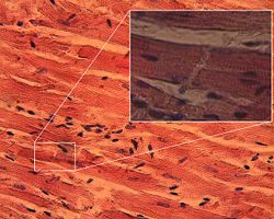
However, cardiac muscle has unique features relative to skeletal muscle. For one, the myocytes are much shorter and are narrower than the skeletal muscle cells, being about 0.1 millimeters long and 0.02 millimeters wide (Ward 2001). Furthermore, while skeletal muscles are arranged in regular, parallel bundles, cardiac muscle connects at branching, irregular angles. Cardiac muscle is anatomically different in that the muscle fibers are typically branched like a tree branch. In addition, cardiac muscle fibers connect to other cardiac muscle fibers through intercalcated discs and form the appearance of a syncytium (continuous cellular material). These intercalcated discs, which appear as irregularly-spaced dark bands between myocytes, are a unique and prominent feature of cardiac muscle (Ward 2001).
On the other hand, cardiac muscle shares many properties with smooth muscle, including being controlled by the autonomic nervous system and spontaneous (automatic) contractions.
Intercalated disc
Intercalated disks are a unique, prominent, and important feature of cardiac muscle. An intercalated disc is an undulating double membrane separating adjacent cells in cardiac muscle fibers. They have two essential functions. For one, they act as a glue to hold myocytes together so that they do not separate when the heart contracts. Secondly, they allow an electrical connection between the cells, supporting synchronized contraction of cardiac tissue. They can easily be visualized by a longitudinal section of the tissue.
Three types of membrane junctions exist within an intercalated disc: fascia adherens, macula adherens, and gap junctions. Fascia adherens are anchoring sites for actin, and connects to the closest sarcomere. Macula adherens stop separation during contraction by binding intermediate filaments joining the cells together, also called a desmosome. Gap junctions contain pores and allow action potentials to spread between cardiac cells by permitting the passage of ions between cells, producing depolarization of the heart muscle.
When observing cardiac tissue through a microscope, intercalated discs are an identifying feature of cardiac muscle
Appearance
Striations. Cardiac muscle exhibits cross striations formed by alternation segments of thick and thin protein filaments, which are anchored by segments called T-lines. The primary structural proteins of cardiac muscle are actin and myosin. The actin filaments are thin causing the lighter appearance of the I bands in muscle, while myosin is thicker and darker lending a darker appearance to the alternating A bands in cardiac muscle as observed by a light enhanced microscope.
T-Tubules. Another histological difference between cardiac muscle and skeletal muscle is that the T-tubules in cardiac muscle are larger, broader, and run along the Z-Discs. There are fewer T-tubules in comparison with skeletal muscle. Additionally, cardiac muscle forms dyads instead of the triads formed between the T-tubules and the sarcoplasmic reticulum in skeletal muscle.
Intercalated discs. Under light microscopy, intercalated discs appear as thin, typically dark-staining lines dividing adjacent cardiac muscle cells. The intercalated discs run perpendicular to the direction of muscle fibers. Under electron microscopy, an intercalated disc's path appears more complex. At low magnification, this may appear as a convoluted electron dense structure overlying the location of the obscured Z-line. At high magnification, the intercalated disc's path appears even more convoluted, with both longitudinal and transverse areas appearing in longitudinal section. Gap junctions (or nexus junctions) fascia adherens (resembling the zonula adherens), and desmosomes are visible. In transverse section, the intercalated disk's appearance is labyrinthine and may include isolated interdigitations.
Contraction mechanism
Cardiac muscle is adapted to be highly resistant to fatigue: it has a large number of mitochondria, enabling continuous aerobic respiration, numerous myoglobins (oxygen-storing pigment), and a good blood supply, which provides nutrients and oxygen. The heart is so tuned to aerobic metabolism that it is unable to pump sufficiently in ischaemic conditions. At basal metabolic rates, about 1% of energy is derived from anaerobic metabolism. This can increase to 10% under moderately hypoxic conditions, but, under more severe hypoxic conditions, not enough energy can be liberated by lactate production to sustain ventricular contractions.[1]
Under basal aerobic conditions, 60% of energy comes from fat (free fatty acids and triacylglycerols/triglycerides), 35% from carbohydrates, and 5% from amino acids and ketone bodies. However, these proportions vary widely according to nutritional state. For example, during starvation, lactate can be recycled by the heart. This is very energy efficient, because one NAD+ is reduced to NADH and H+ (equal to 2.5 or 3 ATP) when lactate is oxidized to pyruvate, which can then be burned aerobically in the TCA cycle, liberating much more energy (ca 14 ATP per cycle).
In the condition of diabetes, more fat and less carbohydrate is used due to the reduced induction of GLUT4 glucose transporters to the cell surfaces. However, contraction itself plays a part in bringing GLUT4 transporters to the surface.[2] This is true of skeletal muscle, but relevant in particular to cardiac muscle, since it is always contracting.
Unlike skeletal muscle, which contracts in response to nerve stimulation, specialized pacemaker cells at the entrance of the right atrium termed the sinoatrial node display the phenomenon of automaticity and are myogenic, meaning that they are self-excitable without a requisite electrical impulse coming from the central nervous system. The rest of the myocardium conducts these action potentials by way of electrical synapses called gap junctions. It is because of this automaticity that an individual's heart does not stop when a neuromuscular blocker (such as succinylcholine or rocuronium) is administered, such as during general anesthesia.
A single cardiac muscle cell, if left without input, will contract rhythmically at a steady rate; if two cardiac muscle cells are in contact, whichever one contracts first will stimulate the other to contract, and so on. This inherent contractile activity is heavily regulated by the autonomic nervous system. If synchronization of cardiac muscle contraction is disrupted for some reason (for example, in a heart attack), uncoordinated contraction known as fibrillation can result.
Rate
Specialized pacemaker cells in the sinoatrial node normally determine the overall rate of contractions, with an average resting pulse of 72 beats per minute.
The central nervous system does not directly create the impulses to contract the heart, but only sends signals to speed up or slow down the heart rate through the autonomic nervous system using two opposing kinds of modulation:
- (1) sympathetic nervous system (fight or flight response)
- (2) parasympathetic nervous system (rest and repose)
Since cardiac muscle is myogenic, the pacemaker serves only to modulate and coordinate contractions. The cardiac muscle cells would still fire in the absence of a functioning SA node pacemaker, albeit in a chaotic and ineffective manner. This condition is known as fibrillation. Note that the heart can still beat properly even if its connections to the central nervous system are completely severed.
Role of calcium
In contrast to skeletal muscle, cardiac muscle cannot contract in the absence of extracellular calcium ions as well as extracellular sodium ions. In this sense, it is intermediate between smooth muscle, which has a poorly developed sarcoplasmic reticulum and derives its calcium across the sarcolemma; and skeletal muscle which is activated by calcium stored in the sarcoplasmic reticulum (SR).
The reason for the calcium dependence is due to the mechanism of calcium-induced calcium release (CICR) from the SR that must occur under normal excitation-contraction (EC) coupling to cause contraction.
ReferencesISBN links support NWE through referral fees
- Indiana State University, Muscle action
- Physiology at MCG 2/2ch7/2ch7line
- Ward, J. 2001. Cardiac muscle. In C. Blakemore, and S. Jennett. The Oxford Companion to the Body. New York: Oxford University Press. ISBN 019852403X
See also
- Myocardium
- Heart
- Circulatory system
- Cardiac action potential
- Calcium sparks
- Troponin
| ||||||||
| |||||||||||
Credits
New World Encyclopedia writers and editors rewrote and completed the Wikipedia article in accordance with New World Encyclopedia standards. This article abides by terms of the Creative Commons CC-by-sa 3.0 License (CC-by-sa), which may be used and disseminated with proper attribution. Credit is due under the terms of this license that can reference both the New World Encyclopedia contributors and the selfless volunteer contributors of the Wikimedia Foundation. To cite this article click here for a list of acceptable citing formats.The history of earlier contributions by wikipedians is accessible to researchers here:
The history of this article since it was imported to New World Encyclopedia:
Note: Some restrictions may apply to use of individual images which are separately licensed.
