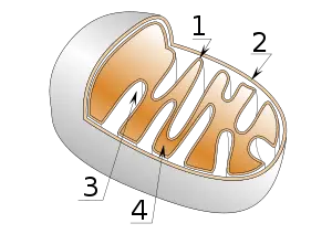
Mitochondrion
A mitochondrion (plural mitochondria) is an organelle found in most eukaryotic cells. Mitochondria are sometimes described as "cellular power plants," because their primary function is to convert organic materials into energy in the form of ATP via the process of oxidative phosphorylation. Usually a cell has hundreds or thousands of mitochondria, which can occupy up to 25 percent of the cell's cytoplasm. The name comes from the Greek mitos, meaning "thread" and khondrion, meaning "granule."
Mitochondria have their own DNA, and, according to the generally accepted endosymbiotic theory, they were originally derived from external organisms. This theory, which was popularized by Lynn Margulis, fits her view that "Life did not take over the globe by combat, but by networking" (Margulis and Sagan 1986)âin other words, by cooperation rather than Darwinian competition.
Mitochondrion structure
A mitochondrion comprises outer and inner membranes composed of phospholipid bilayers studded with proteins, much like a typical cell membrane. The two membranes, however, have very different properties.
The outer mitochondrial membrane, which encloses the entire organelle, comprises by weight about 50 percent phospholipids forming the membranous structure within which float a variety of enzymes involved in such diverse activities as the elongation of fatty acids, oxidation of epinephrine (adrenaline), and the degradation of tryptophan (an essential amino acid). Also floating in the membrane are numerous integral proteins called porins whose relatively large internal channel (about 2-3 nanometers) is permeable to all molecules of 5,000 daltons (a unit of atomic mass) or less (Alberts 1994). Larger molecules can only transverse the outer membrane by active transport (transport aided by a protein and requiring the input of chemical energy).
Unlike the relatively smoothly curved outer membrane, the inner membrane is recursively invaginated, compacting a large membrane surface area into a small volume. In addition to the essential phospholipid foundation needed for forming a biological membrane, the inner membrane also comprises proteins with three types of functions (Alberts 1994):
- Carrying out the oxidation reactions of the respiratory chain.
- Making ATP in the matrix.
- Transporting proteins that regulate the passage of metabolites (intermediates and products of metabolism) into and out of the matrix.
The inner membrane comprises more than one hundred different polypeptides and has a very high protein-to-phospholipid ratio (more than 3:1 by weight, which is about one protein per 15 phospholipids). Additionally, the inner membrane is rich in an unusual phospholipid, cardiolipin, which is usually characteristic of bacterial plasma membranes. Unlike the outer membrane, the inner membrane does not contain porins, and is highly impermeable; almost all ions and molecules require special membrane transporters to enter or exit the matrix.
The mitochondrial matrix
The matrix is the space enclosed by the inner membrane. The matrix contains a highly concentrated mixture of hundreds of enzymes, in addition to the special mitochondrial ribosomes, transfer RNA (tRNA), and several copies of the mitochondrial DNA genome. Of the enzymes, the major functions include oxidation of pyruvate and fatty acids, and the citric acid cycle (Alberts 1994).
Thus, mitochondria possess their own genetic material, and the machinery to manufacture their own RNAs and proteins. This nonchromosomal DNA encodes a small number of mitochondrial peptides (13 in humans) that are integrated into the inner mitochondrial membrane, along with polypeptides encoded by genes that reside in the host cell's nucleus.
Mitochondrial functions
The primary function of mitochondria is to convert organic materials into cellular energy in the form of ATP. Notably, the inner mitochondrial membrane is folded into numerous cristae (see diagram above), which expand the surface area of the inner mitochondrial membrane, enhancing its ability to generate ATP. In typical liver mitochondria, for example, the surface area, including cristae, is about five times that of the outer membrane. Mitochondria of cells that have greater demand for ATP, such as muscle cells, contain even more cristae than typical liver mitochondria.
Mitochondria play an important role in other metabolic tasks:
- Apoptosis (programmed cell death)
- Glutamate-mediated excitotoxic neuronal injury
- Cellular proliferation
- Regulation of the cellular redox state (chemical process in which the oxidation number of atoms is changed)
- Heme synthesis
- Steroid synthesis
- Heat production (enabling the organism to stay warm).
Some mitochondrial functions are performed only in specific types of cells. For example, mitochondria in liver cells contain enzymes that allow them to detoxify ammonia, a waste product of protein metabolism. A mutation in the genes regulating any of these functions can result in a variety of mitochondrial diseases.
Energy conversion
As stated above, the primary function of the mitochondria is the production of ATP. Outside the mitochondria, cells can generate ATP in the absence of oxygen; this process is called glycolysis. Through glycolysis, one molecule of glucose is converted to pyruvate, producing four ATP. Inside the mitochondria, however, much more energy is extracted. This is done by metabolizing the major products of glycolysis: pyruvate and NADH (an important coenzyme, the reduced form of nicotinamide adenine dinucleotide). This metabolism can be performed in two very different ways, depending on the type of cell and the presence or absence of oxygen.
Inside the matrix, the citric acid cycle takes place. The citric acid cycle does not use oxygen. Each pyruvate molecule produced by glycolysis is actively transported across the inner mitochondrial membrane, and into the matrix where it is combined with coenzyme A to form acetyl CoA. Once formed, acetyl CoA is fed into the citric acid cycle , also known as the tricarboxylic acid (TCA) cycle or Krebs cycle. This process creates 3 molecules of NADH and 1 molecule of FADH2, which go on to participate in the next stage, oxidative phosphorylation, which involves oxygen.
The energy from NADH and FADH2 is transferred to oxygen (O2) in several steps via the electron transfer chain. The protein complexes in the inner membrane (NADH dehydrogenase, cytochrome c reductase, cytochrome c oxidase) that perform the transfer use the released energy to pump protons (H+) against a gradient (the concentration of protons in the intermembrane space is higher than that in the matrix).
As the proton concentration increases in the intermembrane space, a strong concentration gradient is built up. The main exit for these protons is through the ATP synthase complex. By transporting protons from the intermembrane space back into the matrix, the ATP synthase complex can make ATP from ADP and inorganic phosphate (Pi). This process is called chemiosmosis and is an example of facilitated diffusion. Peter Mitchell was awarded the 1978 Nobel Prize in Chemistry for his work on chemiosmosis. Later, part of the 1997 Nobel Prize in Chemistry was awarded to Paul D. Boyer and John E. Walker for their clarification of the working mechanism of ATP synthase.
Under certain conditions, protons may be allowed to re-enter the mitochondrial matrix without contributing to ATP synthesis. This process, known as proton leak or mitochondrial uncoupling, results in the unharnessed energy being released as heat. This mechanism for the metabolic generation of heat is employed primarily in specialized tissues, such as the "brown fat" of newborn or hibernating mammals.
The presence of oxygen and the citric acid cycle allows the pyruvate to be broken down into carbon dioxide and water to produce 24-28 ATP.
Reproduction and gene inheritance
Mitochondria replicate their DNA and divide mainly in response to the energy needs of the cellâtheir growth and division is not linked to the cell cycle. When the energy needs of a cell are high, mitochondria grow and divide. When the energy use is low, mitochondria become inactive or are destroyed. During cell division, mitochondria are distributed to the daughter cells more or less randomly during the division of the cytoplasm.
Mitochondria divide by binary fission similar to bacterial cell division. Unlike bacteria, however, mitochondria can also fuse with other mitochondria. Sometimes new mitochondria are synthesized in centers that are rich in proteins and polyribosomes needed for their synthesis.
Mitochondrial genes are not inherited by the same mechanism as nuclear genes. At fertilization of an egg by a sperm, the egg nucleus and sperm nucleus each contributes equally to the genetic makeup of the zygote nucleus. However, all of the mitochondria, and therefore all the mitochondrial genes, are contributed by the egg. At fertilization of an egg, a single sperm enters the egg along with the mitochondria that it uses to provide the energy needed for its swimming behavior. However, the mitochondria provided by the sperm are targeted for destruction very soon after entry into the egg. The egg itself contains relatively few mitochondria, but it is these mitochondria that survive and divide to populate the cells of the adult organism. This type of inheritance is called maternal inheritance and is common to the mitochondria of all animals.
Because mitochondria are inherited from the mother only, the sequence of mitochondrial DNA is sometimes used to trace the lineage of families.
In 1987, Rebecca Cann of the University of Hawaii compared mitochondrial DNA sampled from women whose ancestors came from different parts of the world. The study team compared the differences between the mitochondrial DNA of all the sampled individuals. In this way, they created a family tree connecting them. They used statistical techniques to find a root common to all the women. Africa was determined to be the most likely root of human ancestry.
If the rate of mutation over time could be estimated, they suggested that an approximate date that humans first left Africa could be made. They hypothesized that our human ancestors left Africa between 180,000 and 230,000 years ago.
Origin
As mitochondria contain ribosomes and DNA, and are only formed by the division of other mitochondria, it is generally accepted that they were originally derived from endosymbiotic prokaryotes. Studies of mitochondrial DNA, which is circular and employs a variant genetic code, suggest their ancestor was a member of the Proteobacteria (Futuyma 2005), and probably related to the Rickettsiales.
The endosymbiotic hypothesis suggests that mitochondria descended from specialized bacteria (probably purple nonsulfur bacteria) that somehow survived endocytosis by another species of prokaryote or some other cell type, and became incorporated into the cytoplasm. The ability of symbiont bacteria to conduct cellular respiration in host cells that had relied on glycolysis and fermentation would have provided a considerable evolutionary advantage. Similarly, host cells with symbiotic bacteria capable of photosynthesis would also have an advantage. In both cases, the number of environments in which the cells could survive would have been greatly expanded.
This happened at least two billion years ago and mitochondria still show some signs of their ancient origin. Mitochondrial ribosomes are the 70S (bacterial) type, in contrast to the 80S ribosomes found elsewhere in the cell. As in prokaryotes, there is a very high proportion of coding DNA, and an absence of repeats. Mitochondrial genes are transcribed as multigenic transcripts that are cleaved and polyadenylated to yield mature mRNAs. Unlike their nuclear cousins, mitochondrial genes are small, generally lacking introns (sections of DNA that will be spliced out after transcription, but before the RNA is used), and the chromosomes are circular, conforming to the bacterial pattern.
A few groups of unicellular eukaryotes lack mitochondria: the symbiotic microsporidians, metamonads, and entamoebids, and the free-living pelobionts. While this may suggest that these groups are the most primitive eukaryotes, appearing before the origin of mitochondria, it is now generally held to be an artifactâthat they are descendants of eukaryotes with mitochondria and retain genes or organelles derived from mitochondria. Thus, it appears that there are no primitively amitochondriate eukaryotes, and so the origin of mitochondria may have played a critical part in the development of eukaryotic cells.
ReferencesISBN links support NWE through referral fees
- Alberts, B. et al. 1994. Molecular Biology of the Cell, 3rd Edition. New York: Garland Publishing Inc.
- Cann, R. L., M. Stoneking, and A. C. Wilson. 1987. âMitochondrial DNA and human evolution.â Nature 325: 31-36.
- Futuyma, D. J. 2005. âOn Darwin's Shoulders.â Natural History 114(9):64â68.
- Margulis L. and D. Sagan. 1986. Microcosmos. New York: Summit Books.
- Scheffler, I. E. 2001. âA century of mitochondrial research: Achievements and perspectives.â Mitochondrion 1(1):3â31.
This article contains material from the Science Primer published by the NCBI, which, as a US government publication, is in the public domain at http://www.ncbi.nlm.nih.gov/About/disclaimer.html.
Credits
New World Encyclopedia writers and editors rewrote and completed the Wikipedia article in accordance with New World Encyclopedia standards. This article abides by terms of the Creative Commons CC-by-sa 3.0 License (CC-by-sa), which may be used and disseminated with proper attribution. Credit is due under the terms of this license that can reference both the New World Encyclopedia contributors and the selfless volunteer contributors of the Wikimedia Foundation. To cite this article click here for a list of acceptable citing formats.The history of earlier contributions by wikipedians is accessible to researchers here:
The history of this article since it was imported to New World Encyclopedia:
Note: Some restrictions may apply to use of individual images which are separately licensed.
