Difference between revisions of "Vertebral column" - New World Encyclopedia
Rick Swarts (talk | contribs) |
Rick Swarts (talk | contribs) |
||
| Line 4: | Line 4: | ||
The presence of a vertebral column is one of the key defining characteristics of the subphylum [[Vertebrata]] (animals with backbones), which includes such well-known groups as jawless [[fish]]es, bony fishes, [[shark]]s and [[ray]]s, [[amphibian]]s, [[reptile]]s, [[bird]]s, and [[mammal]]s. However, one group that is commonly placed in Vertebrata is the [[hagfish]]es, which do not have vertebrae. Hagfish have traditionally been classified in [[Agnatha]] (jawless vertebrates) with the [[lamprey]]s, and could represent a degenerate type of vertebrate; however, some taxonomies list them outside Vertebrata. | The presence of a vertebral column is one of the key defining characteristics of the subphylum [[Vertebrata]] (animals with backbones), which includes such well-known groups as jawless [[fish]]es, bony fishes, [[shark]]s and [[ray]]s, [[amphibian]]s, [[reptile]]s, [[bird]]s, and [[mammal]]s. However, one group that is commonly placed in Vertebrata is the [[hagfish]]es, which do not have vertebrae. Hagfish have traditionally been classified in [[Agnatha]] (jawless vertebrates) with the [[lamprey]]s, and could represent a degenerate type of vertebrate; however, some taxonomies list them outside Vertebrata. | ||
| − | All vertebrates also have a [[notochord]]—an internal, flexible, rod-shaped supporting structure—at some point in their life cycle. In higher vertebrates, such as the classes Chondrichthyes (cartilaginous fish), | + | All vertebrates also have a [[notochord]]—an internal, flexible, rod-shaped supporting structure—at some point in their life cycle. In higher vertebrates, such as the classes Chondrichthyes (cartilaginous fish), Mammalia (mammals), and Aves (birds), this notochord is typically present only in the embryonic stages, serving a structural role until the cartilaginous or bony vertebrae form and surround the dorsal nerve cord (although the notochord persists in such primitive fish as [[sturgeon]]s). However, in lampreys and hagfish, the notochord does persist as the main axial support of the body, and in lampreys it persists despite the development of primitive vertebrae made of cartilage. |
==Overview== | ==Overview== | ||
Revision as of 20:56, 25 July 2012

Vertebral column, also known as spinal column, backbone, or spine, is the flexible structure in vertebrates that is formed from cartilaginous or bony structures known as vertebrae and extends from the neck to the tail, protecting the spinal cord, among other functions.
The presence of a vertebral column is one of the key defining characteristics of the subphylum Vertebrata (animals with backbones), which includes such well-known groups as jawless fishes, bony fishes, sharks and rays, amphibians, reptiles, birds, and mammals. However, one group that is commonly placed in Vertebrata is the hagfishes, which do not have vertebrae. Hagfish have traditionally been classified in Agnatha (jawless vertebrates) with the lampreys, and could represent a degenerate type of vertebrate; however, some taxonomies list them outside Vertebrata.
All vertebrates also have a notochord—an internal, flexible, rod-shaped supporting structure—at some point in their life cycle. In higher vertebrates, such as the classes Chondrichthyes (cartilaginous fish), Mammalia (mammals), and Aves (birds), this notochord is typically present only in the embryonic stages, serving a structural role until the cartilaginous or bony vertebrae form and surround the dorsal nerve cord (although the notochord persists in such primitive fish as sturgeons). However, in lampreys and hagfish, the notochord does persist as the main axial support of the body, and in lampreys it persists despite the development of primitive vertebrae made of cartilage.
Overview
In all vertebrates, except for hagfish, the dorsal hollow nerve cord has been surrounded with cartilaginous or bony vertebrae and the notochord generally reduced
Chordata phylum is that they all have, at some time in their life, a notochord, a hollow dorsal nerve cord, and pharyngeal slits. A notochord is an internal, flexible rod that supports the body. Composed of cells derived from the mesoderm, the notochord may be bone or cartilage. In lower vertebrates, it persists throughout life as the main axial support of the body, while in higher vertebrates it is replaced by the vertebral column.
The other living member of Agnatha, the lamprey, has primitive vertebrae made of cartilage.
Overview
rod-shaped supporting structure that is one of the distinguishing features of the phylum Chordatas, being found at some point in the life cycle of all chordates (vertebrates, tunicates, and lancelets). Composed of cells derived from the mesoderm, the notochord defines the primitive axis of the chordate embryo and is retained as the main support in the lancelets. In tunicates, it is only found during the larval stage. Among vertebrates, while the notochord is retained by the adults of the lower vertebrates of class Agnatha (hagfish and lampreys), in higher vertebrates (cartilaginous fish, bony fish, reptiles, amphibians, birds, mammals) it is replaced by the vertebral column.
In vertebrates (subphylum Vertebrata) the notochord becomes surrounded by vertebrae. In the higher vertebrates, Gnathostomata (jawed vertebrates), the notochord is only present in the embryonic stage and is completely replaced by the vertebrae in the adult. This includes Class Chondrichthyes (cartilaginous fish), Class Osteichthyes (bony fish), Class Amphibia (amphibians), Class Reptilia (reptiles), Class Aves (birds), and Class Mammalia (mammals). In members of the vertebrate class Agnatha (jawless fish), the hagfish and lampreys, the notochord remains throughout life and is the first primitive vertebral column.
Lampreys are one of two extant groups of the jawless fish, which are classified as superclass or class Agnatha. While most taxa of Agnatha are extinct, remaining are the hagfish (family Myxinidae of order Myxiniformes of the class Myxini) and the lampreys (family Petromyzontidae of the order Petromyzontiformes of the class Cephalasidomorphi). Both of these lack jaws and both have slimy skin without scales or plates. However, hagfish, which sometimes are known as slime eels, lack vertebrae, while the lampreys, sometimes known as lamprey eels, have primitive vertebrae made of cartilage. Lampreys also have a notochord that remains throughout life, which in most vertebrates is found in the embryo stage only.
They lack bone, do not have paired fins, and barbels are absent (Nelson 1994). They do have a vertebrae made of cartilage and retain the notochord in the adult. (Note that many of these features differ markedly from the other agnathan, the hagfish, which, while it lacks paired fins and bone and retains a notochord, also is characterized by the lack of a vertebrae,
Chordata is a phylum and is broken down into three subphyla: Urochordata, Cephalochordata, and Vertebrata. Members of Urochordata and Cephalochordata live only in the ocean (Towle, 1989). Urochordate larvae have a notochord and a nerve cord but these are lost in adulthood. Cephalochordates have a notochord and a nerve cord but no vertebra. In all vertebrates, except for hagfish, the dorsal hollow nerve cord has been surrounded with cartilaginous or bony vertebrae and the notochord generally reduced. Unlike vertebrates, tunicates and cephalochordates lack any kind of skull. (Those with skulls, that is the vertebrates, are placed in the taxonomic group Craniata.) The dorsal nerve cord in vertebrates develops into a spinal cord with a brain (Towle, 1989).
Over 95 percent of all chordates are vertebrates (Towle, 1989).
Hagfish, while generally classified in Agnatha and in the subphylum Vertebrata, actually lack vertebrae.
The issue is whether the hagfish is itself a degenerate type of vertebrate-fish (most closely related to lampreys), or else may represent a stage which precedes the evolution of the vertebral column (as do lancelets).
Vertebrata is the largest subphylum of the phylum Chordata (chordates), and includes animals with which many people are familiar. Fish (including lampreys), amphibians, reptiles, birds, and mammals (including humans) are vertebrates. Over 50,000 species of vertebrates have been described. However, more than 95 percent of described animal species are invertebrates—a disparate classification of all animals without backbones.
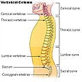
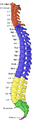
In all animals, vertebrae are defined by the regions of the vertebral column they occur in. Cervical vertebrae are those in the neck area. With exception of two sloth genera (Choleopus and Bradypus) and the manatee (Trichechus), all mammals have seven cervical vertebrae.[1] In other vertebrates it can range from a single vertebra in amphibians, to as many as 25 in swans or 76 in the extinct plesiosaur Elasmosaurus. The dorsal vertebrae range from the bottom of the neck to the top of the pelvis. Dorsal vertebrae attached to ribs are called thoracic vertebrae, while those without ribs are called lumbar vertebrae. The sacral vertebrae are those in the pelvic region, and range from one in amphibians, to two in most birds and modern reptiles, or up to 3 to 5 in mammals. When multiple sacral vertebrae are fused into a single structure, it is called the sacrum. The synsacrum is a similar fused structure found in birds that is composed of the sacral, lumbar, and some of the thoracic and caudal vertebra, as well as the pelvic girdle. Caudal vertebrae compose the tail, and the final few can be fused into the pygostyle in birds, or into the coccygeal or tail bone in chimpanzees (and humans).
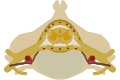
Structure of individual vertebrae
Individual vertebrae are composed of a centrum (body), arches protruding from the top and bottom of the centrum, and various processes projecting from the centrum and/or arches. An arch extending from the top of the centrum is called a neural arch, while the hemal arch or chevron is found underneath the centrum in the caudal (tail) vertebrae of fish, most reptiles, some birds, and some mammals with long tails. The vertebral processes can either give the structure rigidity, help them articulate with ribs, or serve as muscle attachment points. Common types are transverse process, diapophyses, parapophyses, and zygapophyses (both the cranial zygapophyses and the caudal zygapophyses).
Classification
The centra of the vertebra can be classified based upon the fusion of its elements. In aspidospondyly, bones such as the neural spine, the pleurocentrum and the intercentrum are separate ossifications. Fused elements, however, classify a vertebra as having holospondyly.
A vertebra can also be described in terms of the shape of the ends of the centra. Centra with flat ends are acoelous, like those in mammals. These flat ends of the centra are especially good at supporting and distributing compressive forces. Amphicoelous vertebra have centra with both ends concave. This shape is common in fish, where most motion is limited. Amphicoelous centra often are integrated with a full notochord. Procoelous vertebrae are anteriorly concave and posteriorly convex. They are found in frogs and modern reptiles. Opisthocoelous vertebrae are the opposite, possessing anterior convexity and posterior concavity. They are found in salamanders. Heterocoelous vertebrae have saddle-shaped articular surfaces. This type of configuration is seen in turtles that retract their necks, and birds, because it permits extensive lateral and vertical flexion motion without stretching the nerve cord too extensively or wringing it about its long axis.
In non-humans
Fish and amphibians
The vertebrae of lobe-finned fishes consist of three discrete bony elements. The vertebral arch surrounds the spinal cord, and is of broadly similar form to that found in most other vertebrates. Just beneath the arch lies a small plate-like pleurocentrum, which protects the upper surface of the notochord, and below that, a larger arch-shaped intercentrum to protect the lower border. Both of these structures are embedded within a single cylindrical mass of cartilage. A similar arrangement was found in the primitive Labyrinthodonts, but in the evolutionary line that led to reptiles (and hence, also to mammals and birds), the intercentrum became partially or wholly replaced by an enlarged pleurocentrum, which in turn became the bony vertebral body.[2]
In most ray-finned fishes, including all teleosts, these two structures are fused with, and embedded within, a solid piece of bone superficially resembling the vertebral body of mammals. In living amphibians, there is simply a cylindrical piece of bone below the vertebral arch, with no trace of the separate elements present in the early tetrapods.[2]
In cartilagenous fish, such as sharks, the vertebrae consist of two cartilagenous tubes. The upper tube is formed from the vertebral arches, but also includes additional cartilagenous structures filling in the gaps between the vertebrae, and so enclosing the spinal cord in an essentially continuous sheath. The lower tube surrounds the notochord, and has a complex structure, often including multiple layers of calcification.[2]
Lampreys have vertebral arches, but nothing resembling the vertebral bodies found in all higher vertebrates. Even the arches are discontinuous, consisting of separate pieces of arch-shaped cartilage around the spinal cord in most parts of the body, changing to long strips of cartilage above and below in the tail region. Hagfishes lack a true vertebral column, and are therefore not properly considered vertebrates, but a few tiny neural arches are present in the tail.[2]
- Lamprey is the common name for elongated, eel-like, jawless fish comprising the family Petromyzontidae, characterized by a primitive vertebrae made of cartilage. Modern fish are conventionally divided into the jawless fish (about 75 species, including lampreys and hagfish), the cartilaginous fish (about 800 species, including sharks and rays), and the bony fish (with over 26,000 species).
Vertebrates (subphylum Vertebrata) are generally classified into two groups: the Agnatha (jawless vertebrates), and the Gnathostomata (jawed vertebrates). The latter group includes fish with hinged jaws and the tetrapods (amphibians, reptiles, birds, and mammals). Agnatha includes the modern day lampreys (Petromyzontiformes) and hagfish (Myxiniformes) as well as several extinct orders.
Chordata is a phylum and is broken down into three subphyla: Urochordata, Cephalochordata, and Vertebrata. Members of Urochordata and Cephalochordata live only in the ocean (Towle, 1989). Urochordate larvae have a notochord and a nerve cord but these are lost in adulthood. Cephalochordates have a notochord and a nerve cord but no vertebra. In all vertebrates, except for hagfish, the dorsal hollow nerve cord has been surrounded with cartilaginous or bony vertebrae and the notochord generally reduced. Unlike vertebrates, tunicates and cephalochordates lack any kind of skull. (Those with skulls, that is the vertebrates, are placed in the taxonomic group Craniata.) The dorsal nerve cord in vertebrates develops into a spinal cord with a brain (Towle, 1989).
Over 95 percent of all chordates are vertebrates (Towle, 1989). In typical biological classifications, Agnatha and Gnathostomata are each considered a superclass of Vertebrata. However, there are different taxonomies, including ones in which Agnatha is considered a class, or Gnathostomata is not even recognized as a taxon (ITIS 2001), or Agnatha is not recognized (Janvier 1981).
The classification of hagfish has been controversial. The issue is whether the hagfish is itself a degenerate type of vertebrate-fish (most closely related to lampreys), or else may represent a stage which precedes the evolution of the vertebral column (as do lancelets). The original scheme groups hagfish and lampreys together as cyclostomes (or historically, Agnatha), as the oldest surviving clade of vertebrates alongside gnathostomes (the now-ubiquitous jawed-vertebrates). An alternative scheme proposed that jawed-vertebrates are more closely related to lampreys than to hagfish (i.e., that vertebrates include lampreys but exclude hagfish), and introduces the category craniata to group vertebrates near hagfish. Recent DNA evidence has supported the original scheme
Hagfish, while generally classified in Agnatha and in the subphylum Vertebrata, actually lack vertebrae. For this reason, they sometimes are separated from the vertebrates. Janvier (1981) and a number of others, for example, put hagfish into a separate subphylum, Myxini, which then is paired with the subphylum Vertebrata to comprise the taxon Craniata, which recognizes the common possession of a cranium (Janvier 1981). Others, however, use the terms Vertebrata and Craniata as synonyms, rather than as different levels of classification, and retain the use of Agnatha as a superclass (Nelson 1994).
Chordates (phylum Chordata) are a group of animals that includes all the vertebrates (subphylum Vertebrata), as well as two subphylum of invertebrates, the Urochordata (tunicates) and the Cephalochordata (lancelets).
The distinguishing features of the Chordata phylum is that they all have, at some time in their life, a notochord, a hollow dorsal nerve cord, and pharyngeal slits. A notochord is an internal, flexible rod that supports the body. Composed of cells derived from the mesoderm, the notochord may be bone or cartilage. In lower vertebrates, it persists throughout life as the main axial support of the body, while in higher vertebrates it is replaced by the vertebral column.
The other living member of Agnatha, the lamprey, has primitive vertebrae made of cartilage.
Lampreys are one of two extant groups of the jawless fish, which are classified as superclass or class Agnatha. While most taxa of Agnatha are extinct, remaining are the hagfish (family Myxinidae of order Myxiniformes of the class Myxini) and the lampreys (family Petromyzontidae of the order Petromyzontiformes of the class Cephalasidomorphi). Both of these lack jaws and both have slimy skin without scales or plates. However, hagfish, which sometimes are known as slime eels, lack vertebrae, while the lampreys, sometimes known as lamprey eels, have primitive vertebrae made of cartilage. Lampreys also have a notochord that remains throughout life, which in most vertebrates is found in the embryo stage only.
They lack bone, do not have paired fins, and barbels are absent (Nelson 1994). They do have a vertebrae made of cartilage and retain the notochord in the adult. (Note that many of these features differ markedly from the other agnathan, the hagfish, which, while it lacks paired fins and bone and retains a notochord, also is characterized by the lack of a vertebrae,
- Hagfish are the only animals that have a skull but not a vertebral column. Despite their lack of vertebrae, the hagfish have traditionally been classified among the vertebrates.
- Hagfish are jawless and generally classified with the lampreys into the superclass Agnatha (jawless vertebrates) within the subphylum Vertebrata. However, hagfish actually lack vertebrae. For this reason, they sometimes are separated from the vertebrates and not even considered to be fish. Janvier (1981) and a number of others put hagfish in a separate subphylum Myxini, which along with the subphylum Vertebrata comprises the taxon Craniata, recognizing the common possession of a cranium (Janvier 1981). Others, however, place Vertebrata and Craniata as synonyms at the same level of classification, and thereby retain hagfish (Myxini) as members of the superclass Agnatha within the vertebrates (Nelson 1994). The other living member of Agnatha, the lamprey, has primitive vertebrae made of cartilage.
Yet other classifications place Myxini as a class that in one instance lies within the subphylum Vertebrata (ITIS 2003) and, in another instance, lies within—and is the only class in—the clade Craniata, which is considered to be separate from the subphylum Vertebrata (Campbell and Reece 2005).
Campbell, N. A., and J. B. Reece. 2005. Biology, 7th edition. San Francisco: Benjamin Cummings. ISBN 0536964173.
Integrated Taxonomic Information System (ITIS). 2003. Agnatha. ITIS Taxonomic Serial No.: 159693. Retrieved May 31, 2008.
Amniotes
The general structure of human vertebrae is fairly typical of that found in mammals, reptiles, and birds. The shape of the vertebral body does, however, vary somewhat between different groups. In mammals, such as humans, it typically has flat upper and lower surfaces, while in reptiles the anterior surface commonly has a concave socket into which the expanded convex face of the next vertebral body fits. Even these patterns are only generalisations, however, and there may be variation in form of the vertebrae along the length of the spine even within a single species. Some unusual variations include the saddle-shaped sockets between the cervical vertebrae of birds and the presence of a narrow hollow canal running down the centre of the vertebral bodies of geckos and tuataras, containing a remnant of the notochord.[2]
Reptiles often retain the primitive intercentra, which are present as small crescent-shaped bony elements lying between the bodies of adjacent vertebrae; similar structures are often found in the caudal vertebrae of mammals. In the tail, these are attached to chevron-shaped bones called haemal arches, which attach below the base of the spine, and help to support the musculature. These latter bones are probably homologous with the ventral ribs of fish. The number of vertebrae in the spines of reptiles is highly variable, and may be several hundred in some species of snake.[2]
In birds, there is a variable number of cervical vertebrae, which often form the only truly flexible part of the spine. The thoracic vertebrae are partially fused, providing a solid brace for the wings during flight. The sacral vertebrae are fused with the lumbar vertebrae, and some thoracic and caudal vertebrae, to form a single structure, the synsacrum, which is thus of greater relative length than the sacrum of mammals. In living birds, the remaining caudal vertebrae are fused into a further bone, the pygostyle, for attachment of the tail feathers.[2]
Aside from the tail, the number of vertebrae in mammals is generally fairly constant. There are almost always seven cervical vertebrae (sloths and manatees are among the few exceptions), followed by around twenty or so further vertebrae, divided between the thoracic and lumbar forms, depending on the number of ribs. There are generally three to five vertebrae with the sacrum, and anything up to fifty caudal vertebrae.[2]
Human vertebral column
In human anatomy, the vertebral column (Latin − Columna vertebralis) (backbone or spine) is a column usually consisting of 24 articulating vertebrae,[3] and 9 fused vertebrae in the sacrum and the coccyx. It is situated in the dorsal aspect of the torso, separated by intervertebral discs. It houses and protects the spinal cord in its spinal canal.
There are normally thirty-three (33) vertebrae in humans, including the five that are fused to form the sacrum (the others are separated by intervertebral discs) and the four coccygeal bones that form the tailbone. The upper three regions comprise the remaining 24, and are grouped under the names cervical (7 vertebrae), thoracic (12 vertebrae) and lumbar (5 vertebrae), according to the regions they occupy. This number is sometimes increased by an additional vertebra in one region, or it may be diminished in one region, the deficiency often being supplied by an additional vertebra in another. The number of cervical vertebrae is, however, very rarely increased or diminished.[citation needed]

With the exception of the first and second cervical, the true or movable vertebrae (the upper three regions) present certain common characteristics that are best studied by examining one from the middle of the thoracic region.[citation needed]
Structure of individual vertebrae


A typical vertebra consists of two essential parts: an anterior (front) segment, which is the vertebral body; and a posterior part – the vertebral (neural) arch – which encloses the vertebral foramen. The vertebral arch is formed by a pair of pedicles and a pair of laminae, and supports seven processes, four articular, two transverse, and one spinous, the latter also being known as the neural spine.[citation needed]
When the vertebrae are articulated with each other, the bodies form a strong pillar for the support of the head and trunk, and the vertebral foramina constitute a canal for the protection of the medulla spinalis (spinal cord). In between every pair of vertebrae are two apertures, the intervertebral foramina, one on either side, for the transmission of the spinal nerves and vessels.[citation needed]
Two transverse processes and one spinous process are posterior to (behind) the vertebral body. The spinous process comes out the back, one transverse process comes out the left, and one on the right. The spinous processes of the cervical and lumbar regions can be felt through the skin.[citation needed]
Superior and inferior articular facets on each vertebra act to restrict the range of movement possible. These facets are joined by a thin portion of the neural arch called the pars interarticularis.[citation needed]
Curves
Viewed laterally the vertebral column presents several curves, which correspond to the different regions of the column, and are called cervical, thoracic, lumbar, and pelvic.[citation needed]
The cervical curve, convex forward, begins at the apex of the odontoid (tooth-like) process, and ends at the middle of the second thoracic vertebra; it is the least marked of all the curves.[citation needed]
The thoracic curve, concave forward, begins at the middle of the second and ends at the middle of the twelfth thoracic vertebra. Its most prominent point behind corresponds to the spinous process of the seventh thoracic vertebra. This curve is known as a tt curve.[citation needed]
The lumbar curve is more marked in the female than in the male; it begins at the middle of the last thoracic vertebra, and ends at the sacrovertebral angle. It is convex anteriorly, the convexity of the lower three vertebrae being much greater than that of the upper two. This curve is described as a lordotic curve.[citation needed]
The pelvic curve begins at the sacrovertebral articulation, and ends at the point of the coccyx; its concavity is directed downward and forward.[citation needed]
The thoracic and pelvic curves are termed primary curves, because they alone are present during fetal life. The cervical and lumbar curves are compensatory or secondary, and are developed after birth, the former when the child is able to hold up its head (at three or four months) and to sit upright (at nine months), the latter at twelve or eighteen months, when the child begins to walk.[citation needed]
Regions

There are a total of 33 vertebrae in the vertebral column, if assuming 4 coccygeal vertebrae.
The individual vertebrae, named according to region and position, from superior to inferior, are:
- Cervical: 7 vertebrae (C1–C7)
- Thoracic: 12 vertebrae (T1–T12)
- Lumbar: 5 vertebrae (L1–L5)
- Sacral: 5 (fused) vertebrae (S1–S5)
- Coccygeal: 4 (3–5) (fused) vertebrae (Tailbone)
Cervical
There are seven (7) cervical bones (but 8 cervical spinal nerves) and these bones are, in general, small and delicate. Their spinous processes are short (with the exception of C2 and C7, which have palpable spinous processes). Numbered top-to-bottom from C1-C7, atlas (C1) and axis (C2), are the vertebrae that allow the neck and head so much movement. For the most part, the atlanto-occipital joint allows the skull to move up and down, while the atlanto-axial joint allows the upper neck to twist left and right. The axis also sits upon the first intervertebral disk of the spinal column. All mammals except manatees and sloths have seven cervical vertebrae, whatever the length of the neck.[5]
Cervical vertebrae possess transverse foramina to allow for the vertebral arteries to pass through on their way to the foramen magnum to end in the circle of Willis. These are the smallest, lightest vertebrae and the vertebral foramina are triangular in shape. The spinous processes are short and often bifurcated (the spinous process of C7, however, is not bifurcated, and is substantially longer than that of the other cervical spinous processes).[citation needed]
The term cervicothoracic is often used to refer to the cervical and thoracic vertebrae together, and sometimes also their surrounding areas.
Thoracic
The twelve (12) thoracic bones and their transverse processes have surfaces that articulate with the ribs. Some rotation can occur between the thoracic vertebrae, but their connection with the rib cage prevents much flexion or other excursion. They may also be known as 'dorsal vertebrae', in the human context.[citation needed]
Bodies are roughly heart-shaped and are about as wide anterio-posterioly as they are in the transverse dimension. Vertebral foramina are roughly circular in shape.[citation needed]
The term thoracolumbar is sometimes used to refer to the thoracic and lumbar vertebrae together, and sometimes also their surrounding areas.
Lumbar

These five (5) vertebrae are very robust in construction, as they must support more weight than other vertebrae. They allow significant flexion and extension, moderate lateral flexion (sidebending), and a small degree of rotation. The discs between these vertebrae create a lumbar lordosis (curvature that is concave posteriorly) in the human spine.[citation needed]
The term lumbosacral is often used to refer to the lumbar and sacral vertebrae together, and sometimes also their surrounding areas.
Sacral
There are five (5) vertebrae (S1-S5) which are fused in maturity, with no intervertebral discs. The 5 fused bones are collectively known as the sacrum. [6]
Coccygeal
There are usually four (4) and rarely 3 or 5 vertebrae (Co1-Co5), with no intervertebral discs. Many animals have a greater number of "tail vertebrae," and, in animals, they are more commonly known as "caudal vertebrae." Pain at the coccyx (tailbone) is known as coccydynia.[citation needed]
Development
The striking segmented pattern of the human spine is established during embryogenesis when the precursor of the vertebrae, the somites, are rhythmically added to the forming posterior part of the embryo. In humans, somite formation begins around the third week post-fertilization and continues until a total of around 52 somites are formed. The somites are epithelial spheres that contain the precursors of the vertebrae, the ribs, the skeletal muscles of the body wall and limbs, and the dermis of the back. The periodicity of somite distribution and production is thought to be imposed by a molecular oscillator or clock acting in cells of the presomitic mesoderm (PSM). Somites form soon after the beginning of gastrulation, on both sides of the neural tube from a tissue called the presomitic mesoderm (PSM). The PSM is part of the paraxial mesoderm and is generated caudally by gastrulation when cells ingress through the primitive streak, and later, through the tail bud. Soon after their formation, somites become subdivided into the dermomyotome dorsally, which gives rise to the muscles and dermis, and the sclerotome ventrally, which will form the spine components. Sclerotomes become subvidided into an anterior and a posterior compartment. This subdivision plays a key role in the definitive patterning of vertebrae that form when the posterior part of one somite fuses to the anterior part of the consecutive somite during a process termed resegmentation. Disruption of the somitogenesis process in humans results in diseases such as congenital scoliosis. So far, the human homologues of three genes associated to the mouse segmentation clock (MESP2, DLL3 and LFNG) have been shown to be mutated in human patients with human congenital scoliosis suggesting that the mechanisms involved in vertebral segmentation are conserved across vertebrates. In humans the first four somites are incorporated in the basi-occipital bone of the skull and the next 33 somites will form the vertebrae.[7] The remaining posterior somites degenerate. During the fourth week of embryonic development, the sclerotomes shift their position to surround the spinal cord and the notochord. The sclerotome is made of mesoderm and originates from the ventromedial part of the somites. This column of tissue has a segmented appearance, with alternating areas of dense and less dense areas.[citation needed]
As the sclerotome develops, it condenses further eventually developing into the vertebral body. Development of the appropriate shapes of the vertebral bodies is regulated by HOX genes.
The less dense tissue that separates the sclerotome segments develop into the intervertebral discs.
The notochord disappears in the sclerotome (vertebral body) segments, but persists in the region of the intervertebral discs as the nucleus pulposus. The nucleus pulposus and the fibers of the annulus fibrosus make up the intervertebral disc.[citation needed]
The primary curves (thoracic and sacral curvatures) form during fetal development. The secondary curves develop after birth. The cervical curvature forms as a result of lifting the head and the lumbar curvature forms as a result of walking.[citation needed]

There are various defects associated with vertebral development. Scoliosis will result in improper fusion of the vertebrae. In Klippel-Feil anomaly patients have two or more cervical vertebrae that are fused together, along with other associated birth defects. One of the most serious defects is failure of the vertebral arches to fuse. This results in a condition called spina bifida. There are several variations of spina bifida that reflect the severity of the defect.[citation needed]
Surfaces
Anterior surface

When viewed from in front, the width of the bodies of the vertebrae is seen to increase from the second cervical to the first thoracic; there is then a slight diminution in the next three vertebrae; below this there is again a gradual and progressive increase in width as low as the sacrovertebral angle. From this point there is a rapid diminution, to the apex of the coccyx.[citation needed]
Posterior surface

The posterior surface of the vertebral column presents in the median line the spinous processes. In the cervical region (with the exception of the second and seventh vertebrae) these are short and horizontal, with bifid extremities. In the upper part of the thoracic region they are directed obliquely downward; in the middle they are almost vertical, and in the lower part they are nearly horizontal. In the lumbar region they are nearly horizontal. The spinous processes are separated by considerable intervals in the lumbar region, by narrower intervals in the neck, and are closely approximated in the middle of the thoracic region. Occasionally one of these processes deviates a little from the median line — a fact to be remembered in practice, as irregularities of this sort are attendant also on fractures or displacements of the vertebral column. On either side of the spinous processes is the vertebral groove formed by the laminae in the cervical and lumbar regions, where it is shallow, and by the laminae and transverse processes in the thoracic region, where it is deep and broad; these grooves lodge the deep muscles of the back. Lateral to the vertebral grooves are the articular processes, and still more laterally the transverse processes. In the thoracic region, the transverse processes stand backward, on a plane considerably behind that of the same processes in the cervical and lumbar regions. In the cervical region, the transverse processes are placed in front of the articular processes, lateral to the pedicles and between the intervertebral foramina. In the thoracic region they are posterior to the pedicles, intervertebral foramina, and articular processes. In the lumbar region they are in front of the articular processes, but behind the intervertebral foramina.[citation needed]
Lateral surfaces
The lateral surfaces are separated from the posterior surface by the articular processes in the cervical and lumbar regions, and by the transverse processes in the thoracic region. They present, in back, the sides of the bodies of the vertebrae, marked in the thoracic region by the facets for articulation with the heads of the ribs. More posteriorly are the intervertebral foramina, formed by the juxtaposition of the vertebral notches, oval in shape, smallest in the cervical and upper part of the thoracic regions, and gradually increasing in size to the last lumbar. They transmit the special spinal nerves and are situated between the transverse processes in the cervical region, and in front of them in the thoracic and lumbar regions.[citation needed]
Vertebral canal
The vertebral canal follows the different curves of the column; it is large and triangular in those parts of the column which enjoy the greatest freedom of movement, such as the cervical and lumbar regions; and is small and rounded in the thoracic region, where motion is more limited.[citation needed]
Abnormalities
Occasionally the coalescence of the laminae is not completed, and consequently a cleft is left in the arches of the vertebrae, through which a protrusion of the spinal membranes (dura mater and arachnoid), and generally of the spinal cord (medulla spinalis) itself, takes place, constituting the malformation known as spina bifida. This condition is most common in the lumbosacral region, but it may occur in the thoracic or cervical region, or the arches throughout the whole length of the canal may remain incomplete.[citation needed]
The following abnormal curvatures may occur in some people:
- Kyphosis is an exaggerated kyphotic (posterior) curvature in the thoracic region. This produces the so-called "humpback" or "dowager's hump", a condition commonly observed in osteoporosis.
- Lordosis is an exaggerated lordotic (anterior) curvature of the lumbar region, "swayback". Temporary lordosis is common among pregnant women.
- Retrolisthesis is a posterior displacement of one vertebral body with respect to the adjacent vertebral segment to a degree less than a luxation (dislocation).
- Scoliosis, lateral curvature, is the most common abnormal curvature, occurring in 0.5% of the population. It is more common among females and may result from unequal growth of the two sides of one or more vertebrae. It can also be caused by pulmonary atelectasis (partial or complete deflation of one or more lobes of the lungs) as observed in asthma or pneumothorax.
Additional images
Vertebral column.
The spinal cord nested in the vertebral column.
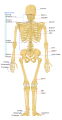
Human skeleton back
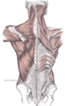
Relation of the vertebral column to the surrounding muscles.
Vertebral column.


ReferencesISBN links support NWE through referral fees
- ↑ Frietson Galis (1999). Why do almost all mammals have seven cervical vertebrae? Developmental constraints, Hox genes and Cancer. Journal of experimental zoology 285:19-26..
- ↑ 2.0 2.1 2.2 2.3 2.4 2.5 2.6 2.7 Romer, Alfred Sherwood (1977). The Vertebrate Body. Philadelphia, PA: Holt-Saunders International, 161–170. ISBN 0-03-910284-X.
- ↑ Template:DorlandsDict
- ↑ Anatomy Compendium (Godfried Roomans and Anca Dragomir)
- ↑ books.google.com
- ↑ Drake et al, Gray's Anatomy for Students, Churchill Livingstone/Elsevier (2010), 2nd edition, chapter 2
- ↑ Somites, Spinal Ganglia, and Centra
- Stemple, D. L. 2005. Structure and function of the notochord: an essential organ for chordate development
June 1, 2005 Development 132, 2503-2512.
Derek L. Stemple
Credits
New World Encyclopedia writers and editors rewrote and completed the Wikipedia article in accordance with New World Encyclopedia standards. This article abides by terms of the Creative Commons CC-by-sa 3.0 License (CC-by-sa), which may be used and disseminated with proper attribution. Credit is due under the terms of this license that can reference both the New World Encyclopedia contributors and the selfless volunteer contributors of the Wikimedia Foundation. To cite this article click here for a list of acceptable citing formats.The history of earlier contributions by wikipedians is accessible to researchers here:
The history of this article since it was imported to New World Encyclopedia:
Note: Some restrictions may apply to use of individual images which are separately licensed.