Difference between revisions of "Intestine" - New World Encyclopedia
Rick Swarts (talk | contribs) |
Rick Swarts (talk | contribs) |
||
| Line 3: | Line 3: | ||
[[Image:Stomach colon rectum diagram.svg|right]] | [[Image:Stomach colon rectum diagram.svg|right]] | ||
| − | In [[anatomy]], the '''intestine''' is | + | In [[anatomy]], the '''intestine''' is that tubular portion of the [[Gastrointestinal tract]] (alimentary canal or digestive tract) of [[vertebrate]]s extending from the [[stomach]] to the [[anus]] or cloaca. The intestine tends to be divided into a [[small intestine]] and a [[large intestine]], with the lower portion designated the large intestine. In humans, the small intestine is further subdivided into the [[duodenum]], [[jejunum]], and [[ileum]], while the large intestine is subdivided into the [[Large intestine#Caecum|cecum]] and colon. |
| + | |||
| + | Although there are huge differences in size and complexity among taxa, in all species the intestine is involved in four functions: digestion and absorption of nutrients, recovery of [[water]] and [[electrolytes]] ([[sodium]], [[chloride]]) from indigestible food matter, formation and storage of feces, and microbial fermentation (Bowen 2006). The small intestine generally also has an immune function in protection against invaders. | ||
| + | |||
| + | ==Diversity among vertebrates== | ||
| + | In cartilaginous [[fish]]es and some primitive bony fishes (eg., lungfish, sturgeon), the intestine is relatively straight and short, and many fishes have a spiral valve (Ritchison 2007). [[Amphibian]]s, [[reptile]]s, [[bird]]s, and [[mammal]]s, as well as some fish, tend to have an elongated and coiled small intestine (Ritchison 2007). In mammals, including humans, the small intestine is divided into three sections: the [[duodenum]], [[jejunum]], and [[ileum]]. Although it is called "small intestine," it is longer in mammals than is the long intestine, but is narrower in diameter. | ||
| + | |||
| + | While the function of the large intestine remains basically the same—absorbing the remaining water and electrolytes from ingesta, forming, storing and eliminating these unusable food matter (wastes), and microbial [[fermentation]]—the size and complexity varies among taxa. Some vertebrate taxa lack a large intestine. For example, killifish ''(Fundulus heteroclitus)'' have a simple digestive system lacking both a large intestine and stomach (but possessing a small intestine) (Vetter et al. 1985) and [[insectivore]]s lack a large intestine (Palaeos 2003). Herbivores like [[horse]]s and [[rabbit]]s, which depend on microbial fermentation, tend to have a very large and complex large intestine, while carnivores like [[cat]]s and [[dog]]s tend to have a simple and small large intestine (Bowen 2000). Omnivores like [[pig]]s and [[human]]s tend to have a substantial large intestine, but smaller and less complex than that of herbivores (Bowen 2000). | ||
| + | |||
| + | Three major portions of the large intestine generally are recognized in [[mammal]]s: ''caecum'' (blind-ended pouch), ''colon'' (majority of the length of the intestine), and ''rectum'' (short, terminal segment) (Bowen 2000). The colon often is incorrectly used in the meaning of the whole large intestine altogether; it is really only the biggest part of the large intestine. | ||
| + | |||
| + | Although called the large intestine, in mammals this tube is shorter than the small intestine, but is wider. | ||
== Structure and Function == | == Structure and Function == | ||
Revision as of 17:39, 17 December 2007
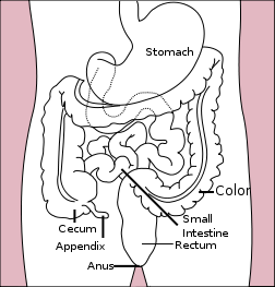
In anatomy, the intestine is that tubular portion of the Gastrointestinal tract (alimentary canal or digestive tract) of vertebrates extending from the stomach to the anus or cloaca. The intestine tends to be divided into a small intestine and a large intestine, with the lower portion designated the large intestine. In humans, the small intestine is further subdivided into the duodenum, jejunum, and ileum, while the large intestine is subdivided into the cecum and colon.
Although there are huge differences in size and complexity among taxa, in all species the intestine is involved in four functions: digestion and absorption of nutrients, recovery of water and electrolytes (sodium, chloride) from indigestible food matter, formation and storage of feces, and microbial fermentation (Bowen 2006). The small intestine generally also has an immune function in protection against invaders.
Diversity among vertebrates
In cartilaginous fishes and some primitive bony fishes (eg., lungfish, sturgeon), the intestine is relatively straight and short, and many fishes have a spiral valve (Ritchison 2007). Amphibians, reptiles, birds, and mammals, as well as some fish, tend to have an elongated and coiled small intestine (Ritchison 2007). In mammals, including humans, the small intestine is divided into three sections: the duodenum, jejunum, and ileum. Although it is called "small intestine," it is longer in mammals than is the long intestine, but is narrower in diameter.
While the function of the large intestine remains basically the same—absorbing the remaining water and electrolytes from ingesta, forming, storing and eliminating these unusable food matter (wastes), and microbial fermentation—the size and complexity varies among taxa. Some vertebrate taxa lack a large intestine. For example, killifish (Fundulus heteroclitus) have a simple digestive system lacking both a large intestine and stomach (but possessing a small intestine) (Vetter et al. 1985) and insectivores lack a large intestine (Palaeos 2003). Herbivores like horses and rabbits, which depend on microbial fermentation, tend to have a very large and complex large intestine, while carnivores like cats and dogs tend to have a simple and small large intestine (Bowen 2000). Omnivores like pigs and humans tend to have a substantial large intestine, but smaller and less complex than that of herbivores (Bowen 2000).
Three major portions of the large intestine generally are recognized in mammals: caecum (blind-ended pouch), colon (majority of the length of the intestine), and rectum (short, terminal segment) (Bowen 2000). The colon often is incorrectly used in the meaning of the whole large intestine altogether; it is really only the biggest part of the large intestine.
Although called the large intestine, in mammals this tube is shorter than the small intestine, but is wider.
Structure and Function
The intestinal tract can be broadly divided into two different parts, the small and large intestine. Grayish-purple in color and about 35 mm (1.5 inches) in diameter, the small intestine is the first and longest, measuring 6-8 meters (22-25 feet) on average in an adult man. Shorter and relatively stockier, the large intestine is a dark reddish color, measuring roughly 1.5 meters (5 feet) on average. Both intestines share a general structure with the whole gut, and is composed of several layers. The lumen is the cavity where digested material passes through and from where nutrients are absorbed. Along the whole length of the gut in the glandular epithelium are goblet cells. These secrete mucus which lubricates the passage of food along and protects it from digestive enzymes. Villi are vaginations of the mucosa and increase the overall surface area of the intestine while also containing a lacteal, which is connected to the lymph system and aids in the removal of lipids and tissue fluid from the blood supply. Micro villi are present on the epithelium of a villus and further increase the surface area over which absorption can take place.
The next layer is the muscularis mucosa which is a layer of smooth muscle that aids in the action of continued peristalsis along the gut. The submucosa contains nerves, blood vessels and elastic fiber with collagen that stretches with increased capacity but maintains the shape of the intestine. Surrounding this is the muscularis externa which comprises longitudinal and smooth muscle that again helps with continued peristalsis and the movement of digested material out of and along the gut.
Lastly there is the serosa which is made up of loose connective tissue and coated in mucus so as to prevent friction damage from the intestine rubbing against other tissue. Holding all this in place are the mesenteries which suspend the intestine in the abdominal cavity and stop it being disturbed when a person is physically active.
The large intestine hosts several kinds of bacteria that deal with molecules the human body is not able to breakdown itself. This is an example of symbiosis. These bacteria also account for the production of gases inside our intestine (this gas is released as flatulence when removed through the anus). However the large intestine is mainly concerned with the absorption of water from digested material (which is regulated by the hypothalamus), the reabsorption of sodium, as well as any nutrients that may have escaped primary digestion in the ileum.
Absorption of glucose in the ileum
Initially, nutrients diffuse passively from the lumen of the ileum via the epithelial cells and into the blood stream. However, certain molecules like glucose passively diffuse in mass quantity some time after a meal, causing a change in concentration gradient. This results in a higher concentration of glucose in the blood (blood sugar level) than in the ileum, such that passive diffusion is no longer possible. Active uptake would be a waste of energy, so another process is used to transport the left-over glucose from the lumen into the blood stream.
In this process, called secondary active transport, a glucose molecule associates with a sodium ion and approaches a transporter protein in the membrane of an epithelial cell. The protein allows the sodium ion through, which then "pulls" the glucose molecule into the cell. Once inside the cell, the sodium and glucose dissociate, and the glucose molecule is free to diffuse passively from the cell into the blood stream (this is because the blood flowing past the cell has a lower blood sugar level than the cell cytoplasm).
Diseases
- Gastroenteritis is inflammation of the intestines and is the most common disease of the intestines. It can arise as the result of food poisoning.
- Ileus is a blockage of the intestines.
- Ileitis is an inflammation of the ileum.
- Colitis is an inflammation of the large intestine.
- Appendicitis is inflammation of the vermiform appendix located at the cecum. This is a potentially fatal disease if left untreated; most cases of appendicitis require surgical intervention.
- Coeliac disease is a common form of malabsorption, affecting up to 1% of people of northern European descent. Allergy to gluten proteins, found in wheat, barley and rye, causes villous atrophy in the small intestine. Life-long dietary avoidance of these foodstuffs in a gluten-free diet is the only treatment.
- Crohn's disease and ulcerative colitis are examples of inflammatory bowel disease. While Crohn's can affect the entire gastrointestinal tract, ulcerative colitis is limited to the large intestine. Crohn's disease is widely regarded as an autoimmune disease. Although ulcerative colitis is often treated as though it were an autoimmune disease, there is no consensus that it actually is such. (See List of autoimmune diseases).
- Enteroviruses are named by their transmission-route through the intestine (enteric = related to intestine), but their symptoms aren't mainly associated with the intestine.
Disorders
- Irritable bowel syndrome is the most common functional disorder of the intestine. Functional constipation and chronic functional abdominal pain are other disorders of the intestine that have physiological causes, but do not have identifiable structural, chemical, or infectious pathologies. They are aberrations of normal bowel function but not diseases.
- Diverticular disease is a condition that is very common in older people in industrialized countries. It usually affects the large intestine but has been known to affect the small intestine as well. Diverticular disease occurs when pouches form on the intestinal wall. Once the pouches become inflamed it is known as Diverticulitis, or Diverticular disease.
- Endometriosis can affect the intestines, with similar symptoms to IBS.
- Bowel twist (or similarly, bowel strangulation) is a comparatively rare event (usually developing sometime after major bowel surgery). It is, however, hard to diagnose correctly, and if left uncorrected can lead to bowel infarction and death. (The singer Maurice Gibb is understood to have died from this.)
See also
- Inflammatory bowel disease (or "IBD")
- Diarrhea
- Constipation
ReferencesISBN links support NWE through referral fees
- Bowen, R. 2006. The large intestine: Introduction and index. Colorado State. Retrieved July 1, 2007.
- Bowen, R. 2000. Gross and microscopic anatomy of the large intestine. Colorado State. Retrieved July 1, 2007.
- Palaeos. 2003. Insectivora. Palaeos. Retrieved July 1, 2007.
- Vetter, R. D., M. C. Carey, and J. S. Patton. 1985. Coassimilation of dietary fat and benzo(a)pyrene in the small intestine: An absorption model using the killifish. Journal of Lipid Research 26: 428-434.
- Ritchison, G. 2007. BIO 342, Comparative Vertebrate Anatomy: Lecture notes 7—Digestive system Gary Ritchison's Home Page, Eastern Kentucky University. Retrieved November 23, 2007.
- Solomon, E. P., L. R. Berg, and D. W. Martin. 2002. Biology. Pacific Grove, CA: Brooks/Cole Thomson Learning. ISBN 0030335035.
- Thomson, A., L. Drozdowski, C. Iodache, B. Thomson, S. Vermeire, M. Clandinin, and G. Wild. 2003. Small bowel review: Normal physiology, part 1. Dig Dis Sci 48(8): 1546-1564. PMID 12924651 Retrieved November 23, 2007.
- Thomson, A., L. Drozdowski, C. Iodache, B. Thomson, S. Vermeire, M. Clandinin, and G. Wild. 2003. Small bowel review: Normal physiology, part 2. Dig Dis Sci 48(8): 1565-1581. PMID 12924652 Retrieved November 23, 2007.
- Townsend, C. M., and D. C. Sabiston. 2004. Sabiston Textbook of Surgery: The Biological Basis of Modern Surgical Practice. Philadelphia: Saunders. ISBN 0721604099.
| Digestive system - edit |
|---|
| Mouth | Pharynx | Esophagus | Stomach | Pancreas | Gallbladder | Liver | Small intestine (duodenum, jejunum, ileum) | Colon | Cecum | Rectum | Anus |
| Endocrine system - edit |
|---|
| Adrenal gland | Corpus luteum | Hypothalamus | Kidney | Ovaries | Pancreas | Parathyroid gland | Pineal gland | Pituitary gland | Testes | Thyroid gland |
Credits
New World Encyclopedia writers and editors rewrote and completed the Wikipedia article in accordance with New World Encyclopedia standards. This article abides by terms of the Creative Commons CC-by-sa 3.0 License (CC-by-sa), which may be used and disseminated with proper attribution. Credit is due under the terms of this license that can reference both the New World Encyclopedia contributors and the selfless volunteer contributors of the Wikimedia Foundation. To cite this article click here for a list of acceptable citing formats.The history of earlier contributions by wikipedians is accessible to researchers here:
The history of this article since it was imported to New World Encyclopedia:
Note: Some restrictions may apply to use of individual images which are separately licensed.
