Meninges
| Meninges | |
|---|---|
 | |
| Meninges of the CNS | |
| Gray's | subject #193 872 |
| Artery | middle meningeal artery, meningeal branches of the ascending pharyngeal artery, accessory meningeal artery, branch of anterior ethmoidal artery, meningeal branches of vertebral artery |
| Nerve | middle meningeal nerve, nervus spinosus |
| MeSH | Meninges |
| Dorlands/Elsevier | m_09/12523818 |
The meninges (singular meninx) is the system of membranes which envelops the central nervous system. The meninges consist of three layers: the dura mater, the arachnoid mater, and the pia mater. The primary function of the meninges and of the cerebrospinal fluid is to protect the central nervous system.
Anatomy
Pia mater
The pia or pia mater is a very delicate membrane. It is the meningeal envelope which firmly adheres to the surface of the brain and spinal cord. As such it follows all the minor contours of the brain (gyri and sulci). It is a very thin membrane composed of fibrous tissue covered on its outer surface by a sheet of flat cells thought to be impermeable to fluid. The pia mater is pierced by blood vessels which travel to the brain and spinal cord, and its capillaries are responsible for nourishing the brain.
- The pia mater (Latin: "tender mother", itself a translation from Arabic) is the delicate innermost layer of the meninges - the membranes surrounding the brain and spinal cord. The thin, mesh-like pia mater closely envelops the entire surface of the brain, running down into the fissures of the cortex. It joins with the ependyma which lines the ventricles to form choroid plexuses that produce cerebrospinal fluid. In the spinal cord, the Pia mater attaches to the Dura mater by the denticular ligaments through the arachnoid membrane. The pia mater is a neural crest derivative.
Arachnoid mater
The middle element of the meninges is the arachnoid mater, so named because of its spider web-like appearance. It provides a cushioning effect for the central nervous system. The arachnoid mater exists as a thin, transparent membrane. It is composed of fibrous tissue and, like the pia mater, is covered by flat cells also thought to be impermeable to fluid. The arachnoid does not follow the convolutions of the surface of the brain and so looks like a loosely fitting sac. In the region of the brain, particularly, a large number of fine filaments called arachnoid trabeculae pass from the arachnoid through the subarachnoid space to blend with the tissue of the pia mater.
The arachnoid and pia mater are sometimes together called the leptomeninges.
- The arachnoid mater is one of the three meninges, the membranes that cover the brain and spinal cord. It is interposed between the two other meninges, the more superficial dura mater and the deeper pia mater, and is separated from the pia mater by the subarachnoid space.
- The delicate, spiderweb-like (therefore the name) arachnoid layer, attached to the inside of the dura, surrounds the brain and spinal cord but does not line the brain down into its sulci (folds). Cerebrospinal fluid flows under this membrane in the subarachnoid space, which is full of the delicate fibres of the arachnoid extending down to attach to the pia mater.
- The portions covering the brain and spinal cord are called arachnoidea encephali and arachnoidea spinalis, respectively.
- The arachnoid and pia mater are sometimes considered as a single structure, the leptomeninx, or the plural version, leptomeninges. (Lepto- from the root meaning thin in Greek). Similarly, the dura in this situation is called the pachymeninx.
- Arachnoid is from a Greek root, and means cob web like. The mater designation (meaning mother in Latin) is borrowed from the dura mater and pia mater, which were Latin translations of Arabic terms. While mater does not technically belong with the arachnoid layer, it has nevertheless been adopted by it for uniformity with the other meninges, and arachnoid mater is currently the Terminologia Anatomica international standard.
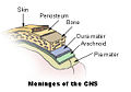
Meninges of the CNS
Diagrammatic section of scalp.

ReferencesISBN links support NWE through referral fees
- Orlando Regional Healthcare, Education and Development. 2004. "Overview of Adult Traumatic Brain Injuries." Retrieved on 2008-01-16.
Dura mater
The dura mater (also rarely called meninx fibrosa, or pachymeninx) is a thick, durable membrane, closest to the skull. It consists of two layers, the periosteal layer, closest to the calvaria and the inner meningeal layer. It contains larger blood vessels which split into the capilliaries in the pia mater. It is composed of dense fibrous tissue, and its inner surface is covered by flattened cells like those present on the surfaces of the pia mater and arachnoid. The dura mater is a sac which envelops the arachnoid and has been modified to serve several functions. The dura mater surrounds and supports the large venous channels (dural sinuses) carrying blood from the brain toward the heart.
Image = Illu meninges.jpg | Caption = Meninges of the CNS | The dura mater (from the Latin "hard mother"), or pachymeninx, is the tough and inflexible outermost of the three layers of the meninges surrounding the brain and spinal cord. (The other two meningeal layers are the pia mater and the arachnoid mater.) The dura mater is not as tightly fitting around the spinal cord, extending past the spinal cord (at the second lumbar vertebra) to about the second sacral vertebra.
Layers and reflections
The dura mater has two layers:
- a superficial layer, which is actually the skull's inner periosteum, called the endocranium.
- a deep layer, the dura mater proper.
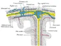
The dura separates into two layers at dural reflections, places where the inner dural layer is reflected as sheet-like protrusions into the cranial cavity. There are two main dural reflections:
- The tentorium cerebelli exists between and separates the cerebellum and brainstem from the occipital lobes of the cerebrum.[1]
- The falx cerebri, which separates the two hemispheres of the brain, is located in the longitudinal cerebral fissure between the hemispheres.[2]
Drainage
The two layers of dura mater run together throughout most of the skull. Where they separate, the gap between them is called a dural venous sinus. These sinuses drain blood and cerebrospinal fluid from the brain and empty into the internal jugular vein.
They drain via the arachnoid villi, which are outgrowths of the arachnoid mater (the middle meningeal layer) that extend into the venous sinuses. These villi act as one-way valves.
Meningeal veins, which course through the dura mater, and bridging veins, which drain the underlying neural tissue and puncture the dura mater, empty into these dural sinuses.
Clinical significance
A subdural hematoma occurs when there is an abnormal collection of blood between the dura and the arachnoid, usually as a result of torn bridging veins secondary to head trauma. An epidural hematoma is a collection of blood between the dura and the inner surface of the skull, and is usually due to arterial bleeding.
The American Red Cross and some other agencies accepting blood donations consider dura mater transplants, along with receipt of pituitary-derived growth hormone, a risk factor due to concerns about Creutzfeldt-Jakob disease.[3]
Dural ectasia is the enlargement of the dura and is common in connective tissue disorders, such as Marfan Syndrome and Ehlers-Danlos Syndrome.
Spaces
The subarachnoid space is the space which normally exists between the arachnoid and the pia mater, which is filled with cerebrospinal fluid.
Normally, the dura mater is attached to the skull, or to the bones of the vertebral canal in the spinal cord. The arachnoid is attached to the dura mater, and the pia mater is attached to the central nervous system tissue. When the dura mater and the arachnoid separate through injury or illness, the space between them is the subdural space.
Pathology
There are three types of hemorrhage involving the meninges:[4]
- A subarachnoid hemorrhage is acute bleeding under the arachnoid; it may occur spontaneously or as a result of trauma.
- A subdural hematoma is a hematoma (collection of blood) located in a separation of the arachnoid from the dura mater. The small veins which connect the dura mater and the arachnoid are torn, usually during an accident, and blood can leak into this area.
- An epidural hematoma similarly may arise after an accident or spontaneously.
Other medical conditions which affect the meninges include meningitis (usually from fungal, bacterial, or viral infection) and meningiomas arising from the meninges or from tumors formed elsewhere in the body which metastasize to the meninges.
Additional images
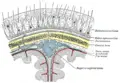
Diagrammatic representation of a section across the top of the skull
Diagrammatic section of scalp.
- Hirnhaut.png

References
- ↑ Shepherd S. 2004. "Head Trauma." Emedicine.com.
- ↑ Vinas FC and Pilitsis J. 2004. "Penetrating Head Trauma." Emedicine.com.
- ↑ Red Cross: http://www.redcross.org/services/biomed/0,1082,0_553_,00.html
- ↑ Orlando Regional Healthcare, Education and Development. 2004. "Overview of Adult Traumatic Brain Injuries." Retrieved on January 16, 2008.
- Blakemore, C., and S. Jennett. 2001. The Oxford Companion to the Body. New York: Oxford University Press. ISBN 019852403X.
- Chamberlin, S. L., and B. Narins. 2005. The Gale Encyclopedia of Neurological Disorders. Detroit: Thomson Gale. ISBN 078769150X.
| |||||||||||
Credits
New World Encyclopedia writers and editors rewrote and completed the Wikipedia article in accordance with New World Encyclopedia standards. This article abides by terms of the Creative Commons CC-by-sa 3.0 License (CC-by-sa), which may be used and disseminated with proper attribution. Credit is due under the terms of this license that can reference both the New World Encyclopedia contributors and the selfless volunteer contributors of the Wikimedia Foundation. To cite this article click here for a list of acceptable citing formats.The history of earlier contributions by wikipedians is accessible to researchers here:
The history of this article since it was imported to New World Encyclopedia:
Note: Some restrictions may apply to use of individual images which are separately licensed.