Difference between revisions of "Bronchus" - New World Encyclopedia
Rick Swarts (talk | contribs) |
Rick Swarts (talk | contribs) |
||
| Line 47: | Line 47: | ||
==Role in disease== | ==Role in disease== | ||
| + | |||
[[Illu_conducting_passages.jpg|thumb|left|240px|Conducting passages]] | [[Illu_conducting_passages.jpg|thumb|left|240px|Conducting passages]] | ||
| + | |||
[[Bronchitis]] is defined as inflammation of the bronchi. There are two main types: acute and chronic. Acute bronchitis is usually caused by viral or bacterial infections. [[Chronic bronchitis]] is a form of [[COPD]], usually associated with smoking or long-term exposure to irritants. | [[Bronchitis]] is defined as inflammation of the bronchi. There are two main types: acute and chronic. Acute bronchitis is usually caused by viral or bacterial infections. [[Chronic bronchitis]] is a form of [[COPD]], usually associated with smoking or long-term exposure to irritants. | ||
[[Asthma]] is hyperreactivity of the bronchi with an inflammatory component, often in response to [[allergen]]s. | [[Asthma]] is hyperreactivity of the bronchi with an inflammatory component, often in response to [[allergen]]s. | ||
Revision as of 22:16, 15 July 2008
| Bronchus | |
|---|---|
 | |
| Conducting passages. | |
 | |
| Front view of cartilages of larynx, trachea, and bronchi. | |
| Gray's | subject #237 1084 |
| System | respiratory |
| Artery | bronchial artery |
| Vein | bronchial vein |
| Nerve | pulmonary branches of vagus nerve |
| MeSH | Bronchi |
| Dorlands/Elsevier | b_23/12198294 |
A bronchus (plural bronchi, adjective bronchial) is a caliber of airway in the respiratory tract that conducts air into the lungs.[1] No gas exchange takes place in this part of the lungs.
Anatomy
The trachea (windpipe) divides into two main bronchi (also mainstem bronchi), the left and the right, at the level of the sternal angle. The right main bronchus is wider, shorter, and more vertical than the left main bronchus. The right main bronchus subdivides into three segmental bronchi while the left main bronchus divides into two. The lobar bronchi divide into tertiary bronchi. Each of the segmental bronchi supplies a bronchopulmonary segment. A bronchopulmonary segment is a division of a lung that is separated from the rest of the lung by a connective tissue septum. This property allows a bronchopulmonary segment to be surgically removed without affecting other segments. There are ten segments per lung, but due to anatomic development, several segmental bronchi in the left lung fuse, giving rise to eight. The segmental bronchi divide into many primary bronchioles which divide into terminal bronchioles, each of which then gives rise to several respiratory bronchioles, which go on to divide into 2 to 11 alveolar ducts. There are 5 or 6 alveolar sacs associated with each alveolar duct. The alveolus is the basic anatomical unit of gas exchange in the lung.
There is hyaline cartilage present in the bronchi, present as irregular rings in the larger bronchi (and not as regular as in the trachea), and as small plates and islands in the smaller bronchi. Smooth muscle is present continuously around the bronchi.
In the mediastinum, at the level of the fifth thoracic vertebra, the trachea divides into the right and left primary bronchi. The bronchi branch into smaller and smaller passageways until they terminate in tiny air sacs called alveoli.
The cartilage and mucous membrane of the primary bronchi are similar to that in the trachea. As the branching continues through the bronchial tree, the amount of hyaline cartilage in the walls decreases until it is absent in the smallest bronchioles. As the cartilage decreases, the amount of smooth muscle increases. The mucous membrane also undergoes a transition from ciliated pseudostratified columnar epithelium to simple cuboidal epithelium to simple squamous epithelium.
The alveolar ducts and alveoli consist primarily of simple squamous epithelium, which permits rapid diffusion of oxygen and carbon dioxide. Exchange of gases between the air in the lungs and the blood in the capillaries occurs across the walls of the alveolar ducts and alveoli.
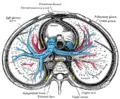
Role in disease
thumb|left|240px|Conducting passages
Bronchitis is defined as inflammation of the bronchi. There are two main types: acute and chronic. Acute bronchitis is usually caused by viral or bacterial infections. Chronic bronchitis is a form of COPD, usually associated with smoking or long-term exposure to irritants. Asthma is hyperreactivity of the bronchi with an inflammatory component, often in response to allergens.
While the left mainstem bronchus departs from the trachea at an angle, the right mainstem bronchus is almost a vertical continuation of the trachea. This anatomy predisposes the right lung to several problems:
- If food, liquids, or foreign bodies are aspirated, they often will lodge in the right mainstem bronchus. Aspiration pneumonia may result.
- If the endotracheal tube used for intubation is inserted too far, it usually lodges in the right mainstem bronchus. This allows ventilation of the right lung, but leaves the left lung useless.
- Patients with inadequate cough reflexes may develop chronic right middle lobe lung infections such as the Lady Windermere Syndrome.
- The right main bronchus (or right primary bronchus, or right principal bronchus) is a bronchus of the respiratory system. It is wider, shorter, and more vertical in direction than the left. It is about 2.5 cm. long, and enters the right lung nearly opposite the fifth thoracic vertebra.
The azygos vein arches over it from behind; and the right pulmonary artery lies at first below and then in front of it.
About 2 cm. from its commencement it gives off a branch to the upper lobe of the right lung.
This is termed the eparterial branch of the bronchus, because it arises above the right pulmonary artery.
The bronchus now passes below the artery, and is known as the hyparterial branch; it divides into two branches for the middle and lower lobes.
Additional images

Transverse section of thorax, showing relations of pulmonary artery.

ReferencesISBN links support NWE through referral fees
- Moore, Keith L. and Arthur F. Dalley. Clinically Oriented Anatomy, 4th ed. (1999). ISBN 0-7817-5936-6
| ||||||||
| ||||||||||||||
Credits
New World Encyclopedia writers and editors rewrote and completed the Wikipedia article in accordance with New World Encyclopedia standards. This article abides by terms of the Creative Commons CC-by-sa 3.0 License (CC-by-sa), which may be used and disseminated with proper attribution. Credit is due under the terms of this license that can reference both the New World Encyclopedia contributors and the selfless volunteer contributors of the Wikimedia Foundation. To cite this article click here for a list of acceptable citing formats.The history of earlier contributions by wikipedians is accessible to researchers here:
The history of this article since it was imported to New World Encyclopedia:
Note: Some restrictions may apply to use of individual images which are separately licensed.
- ↑ Maton, Anthea and Jean Hopkins, Charles William McLaughlin, Susan Johnson, Maryanna Quon Warner, David LaHart, Jill D. Wright (1993). Human Biology and Health. Englewood Cliffs, New Jersey, USA: Prentice Hall. ISBN 0-13-981176-1.