Difference between revisions of "Nerve cord" - New World Encyclopedia
Rick Swarts (talk | contribs) |
Rick Swarts (talk | contribs) |
||
| Line 7: | Line 7: | ||
==Ventral nerve cord== | ==Ventral nerve cord== | ||
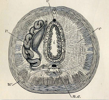
[[File:Anatomy of an earthworm 001.png|thumb|225px|Cross section of an [[earthworm]] showing the central nerve cord (n.c.), as well as the body wall (w); membranes (c) that divide the body cavity into a series of chambers; a coiled nephridium (n); the intestine (i); and above and below it a longer blood vessel (v).]] | [[File:Anatomy of an earthworm 001.png|thumb|225px|Cross section of an [[earthworm]] showing the central nerve cord (n.c.), as well as the body wall (w); membranes (c) that divide the body cavity into a series of chambers; a coiled nephridium (n); the intestine (i); and above and below it a longer blood vessel (v).]] | ||
| − | The '''ventral nerve cord''' is a bundle of nerve fibers, typically a solid double stand (pair) of nerve cords, that runs along the longitudinal axis of some [[phylum|phyla]] of elongate [[invertebrate]]s, and forms part of the invertebrate's [[central nerve system]]. | + | The '''ventral nerve cord''' is a bundle of nerve fibers, typically a solid double stand (pair) of nerve cords, that runs along the longitudinal axis of some [[phylum|phyla]] of elongate [[invertebrate]]s, and forms part of the invertebrate's [[central nerve system]]. In most cases, this nerve cords runs ventrally, below the gut, and connects to the cerebral ganglia. Among the phyla exhibiting ventral nerve cords are [[nematode]]s (roundworms), [[annelid]]s (such as [[earthworm]]s, and [[arthropod]]s (such as [[insect]]s and [[crafish]]). |
The ventral nerve cord usually consists of a pair ''connectives'', which are partially fused nerve trunks running longitudinally along the ventral plane of the animals, from the [[anterior]] to [[Posterior (anatomy)|posterior]] (the [[thorax|thoracic]] and [[abdomen|abdominal]] [[Tagma (arthropod anatomy)|tagma]] in the arthropods). Each body segment is innervated by pairs of ganglia. The segmented ganglia are connected by a tract of nerve fibers passing from one side to the other of the nerve cord called commissures. | The ventral nerve cord usually consists of a pair ''connectives'', which are partially fused nerve trunks running longitudinally along the ventral plane of the animals, from the [[anterior]] to [[Posterior (anatomy)|posterior]] (the [[thorax|thoracic]] and [[abdomen|abdominal]] [[Tagma (arthropod anatomy)|tagma]] in the arthropods). Each body segment is innervated by pairs of ganglia. The segmented ganglia are connected by a tract of nerve fibers passing from one side to the other of the nerve cord called commissures. | ||
| − | There are different degrees of fusion. The complete system bears some likeness to a rope [[ladder]]. In some animals, the bilateral ganglia are fused into a single large ganglion per segment. This characteristic is found mostly in the [[insect]]s. | + | There are different degrees of fusion of the ganglia among different [[taxon]]. The complete system bears some likeness to a rope [[ladder]]. In some animals, the bilateral ganglia are fused into a single large ganglion per segment. This characteristic is found mostly in the [[insect]]s. |
| + | |||
| + | Unlike with [[chordate]]s, the nerve cord in invertebrates does not develop by invagination. Rather than the cells gathering dorsally on the embryo's outer surface, folding inward, and then sinking to their final position, in the case of the ventral nerve cord's formation, the cells commonly move inward to the internal position individually (source??). | ||
==Dorsal nerve cord== | ==Dorsal nerve cord== | ||
| − | The '''dorsal nerve cord''' is | + | The '''dorsal nerve cord''' is a hollow bundle of nerve fibers that transverse dorsally the longitudinal axis of [[chordate]]s, at some stage of their life, and runs above the [[notochord]] and [[gut]]. The dorsal nerve cord is an embryonic feature that is unique to chordates. Other distinguishing features of the Chordata phylum is that they all have, at some stage in their life, a [[notochord]], a [[post-anal tail]], an [[endostyle]], and [[pharyngeal slit]]s. In [[vertebrate]]s, this embryonic feature transforms into the [[brain]] and [[spinal cord]]. |
| − | |||
| − | |||
| − | |||
| − | |||
| − | |||
| − | |||
| − | |||
| − | |||
| − | |||
| − | [[ | + | The dorsal nerve cord develops from a plate of dorsal [[germ layer#Ectoderm|ectoderm]] that invaginates into a hollow, fluid-filled tube. Essentially, the neural tissue, which concentrates above the developing [[notochord]] on the embryo's outer surface, folds into the hollow, neural tube, and then sinks to arrive at its internal position. (***** source?) |
| − | |||
| − | |||
| − | |||
| − | |||
| − | |||
| − | |||
| − | |||
| − | |||
| − | |||
| − | |||
| − | |||
| − | |||
| − | |||
| − | |||
*{{cite book |last=Hickman |first=Cleveland |coauthors=Roberts L. Keen S. Larson A. Eisenhour D|title=Animal Diversity |edition=4th |publisher=McGraw Hill |location=New York|isbn=978-0-07-252844-2}} | *{{cite book |last=Hickman |first=Cleveland |coauthors=Roberts L. Keen S. Larson A. Eisenhour D|title=Animal Diversity |edition=4th |publisher=McGraw Hill |location=New York|isbn=978-0-07-252844-2}} | ||
| Line 48: | Line 27: | ||
* [http://www.lobsters.org/tlcbio/biology6.html Nervous system of a lobster] | * [http://www.lobsters.org/tlcbio/biology6.html Nervous system of a lobster] | ||
* [http://www.entomology.umn.edu/cues/4015/morpology/ Insect morphology] | * [http://www.entomology.umn.edu/cues/4015/morpology/ Insect morphology] | ||
| − | |||
| − | |||
| − | |||
| − | |||
| − | |||
| Line 58: | Line 32: | ||
[[Category: Life sciences]] | [[Category: Life sciences]] | ||
[[Category:Anatomy and physiology]] | [[Category:Anatomy and physiology]] | ||
| + | |||
{{credit|Nerve_cord|543763994|Ventral_nerve_cord|541322392|Dorsal_nerve_cord|553585916}} | {{credit|Nerve_cord|543763994|Ventral_nerve_cord|541322392|Dorsal_nerve_cord|553585916}} | ||
Revision as of 22:19, 2 July 2013
Nerve cord is a term that can refer to either (1) the single, hollow, fluid-filled, dorsal tract of nervous tissue that is one of the defining characteristics of chordates (dorsal nerve cord) and develops into the spinal cord and brain of vertebrates; or (2) the typically solid, ventral, double row of nerve fibers found in some phyla of invertebrates (ventral nerve cord).
In both cases, the term nerve cord references a bundle of nerve fibers that transverse the longitudinal axis of an animal and is an important structure of the animal's central nervous system. However, in the case of chordates, the nerve cord is tubular, hollow, fluid-filled, and runs dorsally, above the notochord and gut tract, while in the case of non-chordates it is solid and runs ventrally, below the digestive tract. They also differ in the the nerve cord of chordates forms by invagination in the embryo, whereas in non-chordates, the nerve cord does not form by invagination.
Ventral nerve cord

The ventral nerve cord is a bundle of nerve fibers, typically a solid double stand (pair) of nerve cords, that runs along the longitudinal axis of some phyla of elongate invertebrates, and forms part of the invertebrate's central nerve system. In most cases, this nerve cords runs ventrally, below the gut, and connects to the cerebral ganglia. Among the phyla exhibiting ventral nerve cords are nematodes (roundworms), annelids (such as earthworms, and arthropods (such as insects and crafish).
The ventral nerve cord usually consists of a pair connectives, which are partially fused nerve trunks running longitudinally along the ventral plane of the animals, from the anterior to posterior (the thoracic and abdominal tagma in the arthropods). Each body segment is innervated by pairs of ganglia. The segmented ganglia are connected by a tract of nerve fibers passing from one side to the other of the nerve cord called commissures.
There are different degrees of fusion of the ganglia among different taxon. The complete system bears some likeness to a rope ladder. In some animals, the bilateral ganglia are fused into a single large ganglion per segment. This characteristic is found mostly in the insects.
Unlike with chordates, the nerve cord in invertebrates does not develop by invagination. Rather than the cells gathering dorsally on the embryo's outer surface, folding inward, and then sinking to their final position, in the case of the ventral nerve cord's formation, the cells commonly move inward to the internal position individually (source??).
Dorsal nerve cord
The dorsal nerve cord is a hollow bundle of nerve fibers that transverse dorsally the longitudinal axis of chordates, at some stage of their life, and runs above the notochord and gut. The dorsal nerve cord is an embryonic feature that is unique to chordates. Other distinguishing features of the Chordata phylum is that they all have, at some stage in their life, a notochord, a post-anal tail, an endostyle, and pharyngeal slits. In vertebrates, this embryonic feature transforms into the brain and spinal cord.
The dorsal nerve cord develops from a plate of dorsal ectoderm that invaginates into a hollow, fluid-filled tube. Essentially, the neural tissue, which concentrates above the developing notochord on the embryo's outer surface, folds into the hollow, neural tube, and then sinks to arrive at its internal position. (***** source?)
- Hickman, Cleveland and Roberts L. Keen S. Larson A. Eisenhour D. Animal Diversity, 4th, New York: McGraw Hill. ISBN 978-0-07-252844-2.
External links
Credits
New World Encyclopedia writers and editors rewrote and completed the Wikipedia article in accordance with New World Encyclopedia standards. This article abides by terms of the Creative Commons CC-by-sa 3.0 License (CC-by-sa), which may be used and disseminated with proper attribution. Credit is due under the terms of this license that can reference both the New World Encyclopedia contributors and the selfless volunteer contributors of the Wikimedia Foundation. To cite this article click here for a list of acceptable citing formats.The history of earlier contributions by wikipedians is accessible to researchers here:
The history of this article since it was imported to New World Encyclopedia:
Note: Some restrictions may apply to use of individual images which are separately licensed.
