Cell membrane
A cell membrane, plasma membrane or plasmalemma is a selectively permeable bilayer which comprises the outer layer of a cell. It is comprised of, among other components, phospholipid and protein molecules which separate the cell interior from its surroundings within animal cells, and control the input and output of cell through the use of receptor and cell adhesion proteins, which also play a role in cell behavior and the organization of cells within tissues.
In animal cells, the cell membrane establishes this separation alone, whereas in yeast, bacteria and plants an additional cell wall forms the outermost boundary, providing primarily mechanical support. The plasma membrane is only about 10 nm thick and may be discerned only faintly with a transmission electron microscope. One of the key roles of the membrane is to maintain the cell potential.
Structure - fluid mosaic
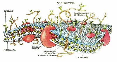
The basic composition and structure of the plasma membrane is the same as that of the membranes that surround organelles and other subcellular compartments. The foundation is a phospholipid bilayer, and the membrane as a whole is often described as a fluid mosaic – a two-dimensional fluid of freely diffusing lipids, dotted or embedded with proteins, which may function as channels or transporters across the membrane, or as receptors. The model was first proposed by S.J. Singer (1971) as a lipid protein model and extended to include the fluid character in a publication with G.L. Nicolson in "Science" (1972).
Some of these proteins simply adhere to the membrane (extrinsic or peripheral proteins), whereas others may be said to reside within it or to span it (intrinsic proteins – more at integral membrane protein). Glycoproteins have carbohydrates attached to their extracellular domains. Cells may vary the variety and the relative amounts of different lipids to maintain the fluidity of their membranes despite changes in temperature. The regulation is assisted the bilayer by cholesterol molecules in eukaryotes and hopanoids in prokaryotes.
Detailed Structure
Phospholipid molecules in the cell membrane are "fluid," in the sense that they are free to diffuse and exhibit rapid lateral diffusion. Lipid rafts and caveolae are examples of cholesterol-enriched microdomains in the cell membrane.
Many proteins are not free to diffuse. The cytoskeleton undergirds the cell membrane and provides anchoring points for integral membrane proteins. Anchoring restricts them to a particular cell face or surface – for example, the "apical" surface of epithelial cells that line the vertebrate gut – and limits how far they may diffuse within the bilayer. Rather than presenting always a formless and fluid contour, the plasma membrane surface of cells may show structure. Returning to the example of epithelial cells in the gut, the apical surfaces of many such cells are dense with involutions, all similar in size. The finger-like projections, called microvilli, increase cell surface area and facilitate the absorption of molecules from the outside. Synapses are another example of highly-structured membrane.
New material is incorporated into the membrane, or deleted from it, by a variety of mechanisms.
- Fusion of intracellular vesicles with the membrane not only excretes the contents of the vesicle, but also incorporates the vesicle membrane's components into the cell membrane. The membrane may form blebs that pinch off to become vesicles.
- If a membrane is continuous with a tubular structure made of membrane material, then material from the tube can be drawn into the membrane continuously.
- Although the concentration of membrane components in the aqueous phase is low (stable membrane components have low solubility in water), exchange of molecules with this small reservoir is possible.
In all cases, the mechanical tension in the membrane has an effect on the rate of exchange. In some cells, usually having a smooth shape, the membrane tension and area are interrelated by elastic and dynamical mechanical properties, and the time-dependent interrelation is sometimes called homeostasis, area regulation or tension regulation.
Structure:
The outer cell membrane and the membranes surrounding inner cell organelles are bilipid layers. To perform the function of the organelle, the membrane is specialized in that it contains specific proteins and lipid components that enable it to perform its unique roles for that cell or organelle. In a the cell membrane phospholipid molecules create a spherical three dimensional lipid bilayer shell around the cell. A phospholipid molecule is composed of a head and two tails. The circle, or head, is the negatively charged phosphate group and the two tails are the two highly hydrophobic fatty acid chains of the phospholipid.
Functions:
1. It attaches parts of the cytoskeleton to the cell membrane in order to provide shape.
2. It attaches cells to an extra-cellular matrix in grouping cells together to form tissues.
3. It transports molecules into and out of cells by such methods as ion pumps, channel proteins and carrier proteins.
4. It acts as receptor for the various chemical messages which pass between cells such as nerve impulses and hormone activity.
5. It takes part in enzyme activity which can be important in the metabolism or as part of the body's defense mechanism.
Transport across membranes
As a lipid bilayer, the cell membrane is semi-permeable. This means that only some molecules can pass unhindered in or out of the cell. These molecules are either small or lipophilic. Other molecules can pass if there are specific transport molecules.
Depending on the molecule, transport occurs by different mechanisms, which can be separated into those that do not consume energy in the form of ATP (passive transport) and those that do (active transport).
Passive transport
Passive transport is a means of moving different chemical substances across membranes through diffusion of hydrophobic (non-polar) and small polar molecules, or facilitated diffusion of polar and ionic molecules, which relies on a transport protein to provide a channel or bind to specific molecules. This spontaneous process decreases free energy, and increases entropy in a system. Unlike active transport, this process does not involve any chemical energy (from ATP).
The outer cell membrane and the membranes surrounding inner cell organelles are bilipid layers. To perform the function of the organelle, the membrane is specialized in that it contains specific proteins and lipid components that enable it to perform its unique roles for that cell or organelle. In the cell membrane phospholipid molecules create a spherical three dimensional lipid bilayer shell around the cell. A phospholipid molecule is composed of a head and two tails. The circle, or head, is the negatively charged phosphate group and the two tails are the two highly hydrophobic fatty acid chains of the phospholipid.
Active transport
Typically moves molecules against their electrochemical gradient, a process that would be entropically unfavorable were it not stoichiometrically coupled with the hydrolysis of ATP. This coupling can be either primary or secondary. In the primary active transport, transporters that move molecules against their electrical/chemical gradient, hydrolyze ATP. In the secondary active transport, transporters use energy derived from transport of another molecule in the direction of their gradient, to move other molecules in the direction against their gradient. This can be either symport (in the same direction) or antiport (in the opposite direction).
Examples include:
- The usual cases of molecular exchangers, transporters and pumps
- endocytosis and exocytosis, where molecules packaged in membrane vesicles are either imported or exported respectively, can be thought of as active.
See also
- Bacterial cell structure
External links
- Lipids, Membranes and Vesicle Trafficking - The Virtual Library of Biochemistry and Cell Biology
- Cell membrane protein extraction protocol
- Membrane homeostasis, tension regulation, mechanosensitive membrane exchange and membrane traffic
| Organelles of the cell |
|---|
| Acrosome | Chloroplast | Cilium/Flagellum | Centriole | Endoplasmic reticulum | Golgi apparatus | Lysosome | Melanosome | Mitochondrion | Myofibril | Nucleus | Parenthesome | Peroxisome | Plastid | Ribosome | Vacuole | Vesicle |
Credits
New World Encyclopedia writers and editors rewrote and completed the Wikipedia article in accordance with New World Encyclopedia standards. This article abides by terms of the Creative Commons CC-by-sa 3.0 License (CC-by-sa), which may be used and disseminated with proper attribution. Credit is due under the terms of this license that can reference both the New World Encyclopedia contributors and the selfless volunteer contributors of the Wikimedia Foundation. To cite this article click here for a list of acceptable citing formats.The history of earlier contributions by wikipedians is accessible to researchers here:
The history of this article since it was imported to New World Encyclopedia:
Note: Some restrictions may apply to use of individual images which are separately licensed.
