Difference between revisions of "Ligament" - New World Encyclopedia
Rick Swarts (talk | contribs) |
Rick Swarts (talk | contribs) |
||
| Line 1: | Line 1: | ||
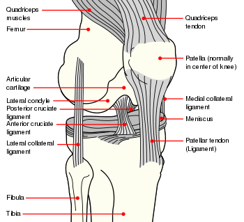
[[Image:Knee diagram.svg|thumb|right|350px|Diagram of the right knee of a human with some of the region's ligaments]]. | [[Image:Knee diagram.svg|thumb|right|350px|Diagram of the right knee of a human with some of the region's ligaments]]. | ||
| − | In [[anatomy]], a '''ligament''' is a band or sheet of strong fibrous [[connective tissue]] that connects bones to other bones, or to cartilage, or supports an organ, such as the [[spleen]], uterus, or eyeball. | + | In [[anatomy]], a '''ligament''' is a band or sheet of strong fibrous [[connective tissue]] that connects bones to other bones, or to cartilage, or supports an organ, such as the [[spleen]], uterus, or eyeball. Such structures tend to be somewhat flexible but inelastic. They differ from [[tendon]]s in that tendons connect skeletal muscles to their attachments. |
| − | The term ligament also is used to denote | + | There are three basic types of structures known as ligaments. Those known as articular ligaments, fibrous ligaments, or true ligaments, connect bones to other bones, such as the anterior cruciate ligament (ACL) that connects the posterio-lateral part of the [[femur]] to an anterio-medial part of the [[tibia]]. The term ligament also is used to denote a fold fold of [[peritoneum]] or other membrane that connects organs, such as the broad ligament of the uterus, which is a wide fold of peritoneum that connects the sides of the [[uterus]] to the walls and floor of the pelvis. In addition, ligament is used to refer to remnants of a tubular structure from the [[fetus|fetal]] period of life, such as the medial umbilical ligament, which is found on the deep surface of the anterior abdominal wall and represents the remnant of the fetal umbilical arteries, but which serves no purpose in humans after birth and is shriveled in adults. |
| + | |||
| + | This article will only briefly review these later two types of ligaments, with the article mainly focusing on the first meaning, which is what is most commonly meant by the term ligament. | ||
The study of ligaments is known as desmology, from the [[Ancient Greek|Greek]] ''desmos'' (δεσμός), meaning "string," and ''-logia'' (-λογία), meaning "study of." | The study of ligaments is known as desmology, from the [[Ancient Greek|Greek]] ''desmos'' (δεσμός), meaning "string," and ''-logia'' (-λογία), meaning "study of." | ||
| − | |||
==Articular ligaments== | ==Articular ligaments== | ||
[[Image:Gray298.png|thumb|Diagrammatic section of a [[symphysis]].]] | [[Image:Gray298.png|thumb|Diagrammatic section of a [[symphysis]].]] | ||
| − | In its most common use, a ''ligament'' is a short band of tough fibrous dense regular [[connective tissue]] composed mainly of long, stringy [[collagen]] [[ | + | In its most common use, a ''ligament'' is a short band of tough, fibrous, dense, regular [[connective tissue]] composed mainly of long, stringy [[collagen]] [[fiber]]. Ligaments commonly connect bones to other bones to form a [[joint]]. They do ''not'' connect [[muscle]]s to bones; that is the function of [[tendon]]s (Blakemore and Jennett 2001). They also |
| + | |||
| + | |||
| + | Some ligaments limit the mobility of articulations, or prevent certain movements altogether. | ||
Capsular ligaments are part of the articular capsule that surrounds synovial [[joints]]. They act as mechanical reinforcements. Extra-capsular ligaments join bones together and provide [[joint]] stability. | Capsular ligaments are part of the articular capsule that surrounds synovial [[joints]]. They act as mechanical reinforcements. Extra-capsular ligaments join bones together and provide [[joint]] stability. | ||
Revision as of 23:23, 16 February 2009
.
In anatomy, a ligament is a band or sheet of strong fibrous connective tissue that connects bones to other bones, or to cartilage, or supports an organ, such as the spleen, uterus, or eyeball. Such structures tend to be somewhat flexible but inelastic. They differ from tendons in that tendons connect skeletal muscles to their attachments.
There are three basic types of structures known as ligaments. Those known as articular ligaments, fibrous ligaments, or true ligaments, connect bones to other bones, such as the anterior cruciate ligament (ACL) that connects the posterio-lateral part of the femur to an anterio-medial part of the tibia. The term ligament also is used to denote a fold fold of peritoneum or other membrane that connects organs, such as the broad ligament of the uterus, which is a wide fold of peritoneum that connects the sides of the uterus to the walls and floor of the pelvis. In addition, ligament is used to refer to remnants of a tubular structure from the fetal period of life, such as the medial umbilical ligament, which is found on the deep surface of the anterior abdominal wall and represents the remnant of the fetal umbilical arteries, but which serves no purpose in humans after birth and is shriveled in adults.
This article will only briefly review these later two types of ligaments, with the article mainly focusing on the first meaning, which is what is most commonly meant by the term ligament.
The study of ligaments is known as desmology, from the Greek desmos (δεσμός), meaning "string," and -logia (-λογία), meaning "study of."
Articular ligaments
In its most common use, a ligament is a short band of tough, fibrous, dense, regular connective tissue composed mainly of long, stringy collagen fiber. Ligaments commonly connect bones to other bones to form a joint. They do not connect muscles to bones; that is the function of tendons (Blakemore and Jennett 2001). They also
Some ligaments limit the mobility of articulations, or prevent certain movements altogether.
Capsular ligaments are part of the articular capsule that surrounds synovial joints. They act as mechanical reinforcements. Extra-capsular ligaments join bones together and provide joint stability.
Ligaments are only elastic; when under tension, they gradually lengthen. (Unlike tendons which are inelastic). This is one reason why dislocated joints must be set as quickly as possible: if the ligaments lengthen too much, then the joint will be weakened, becoming prone to future dislocations. Athletes, gymnasts, dancers, and martial artists perform stretching exercises to lengthen their ligaments, making their joints more supple. The term double-jointed refers to people who have more elastic ligaments, allowing their joints to stretch and contort further. The medical term for describing such double-jointed persons is hyperlaxity and double-jointed is a synonym of hyperlax.
The consequence of a broken ligament can be instability of the joint. Not all broken ligaments need surgery, but if surgery is needed to stabilise the joint, the broken ligament can be joined. Scar tissue may prevent this. If it is not possible to fix the broken ligament, other procedures such as the Brunelli Procedure can correct the instability. Instability of a joint can over time lead to wear of the cartilage and eventually to osteoarthritis.
Examples:
Knee
- Anterior cruciate ligament (ACL)
- Lateral collateral ligament (LCL)
- Posterior cruciate ligament (PCL)
- Medial collateral ligament (MCL)
- Cranial cruciate ligament (CrCL) - quadruped equivalent of ACL
- Caudal cruciate ligament (CaCL) - quadruped equivalent of PCL
Head and neck
- Cricothyroid ligament
- Periodontal ligament
- Suspensory ligament of the lens
Pelvis
- Anterior sacroiliac ligament
- Posterior sacroiliac ligament
- Sacrotuberous ligament
- Sacrospinous ligament
- Inferior pubic ligament
- Superior pubic ligament
- Suspensory ligament of the penis
Thorax
- Suspensory ligament of the breast
Wrist
- See Wrist#Ligaments
See also
- Joint
- Brostrom procedure
Peritoneal ligaments
Certain folds of peritoneum are referred to as ligaments.
Examples include:
- The hepatoduodenal ligament surrounds the hepatic portal vein and other vessels as they travel from the duodenum to the liver.
- The broad ligament of the uterus is also a fold of peritoneum.
- The suspensory ligament of the ovary
Fetal remnant ligaments
Certain tubular structures from the fetal period are referred to as ligaments after they close up and turn into cord-like structures:
| Fetal | Adult |
| ductus arteriosus | ligamentum arteriosum |
| extra-hepatic portion of the fetal left umbilical vein | ligamentum teres hepatis (the "round ligament of the liver"). |
| intra-hepatic portion of the fetal left umbilical vein (the ductus venosus) | ligamentum venosum |
| distal portions of the fetal left and right umbilical arteries | medial umbilical ligaments |
ReferencesISBN links support NWE through referral fees
- Blakemore, C., and S. Jennett. 2001. The Oxford Companion to the Body. New York: Oxford University Press. ISBN 019852403X.
Dorland, William Alexander Newman. 2007. Dorland's illustrated medical dictionary. Edinburgh: Elsevier Saunders. ISBN 9781416023647
http://www.mercksource.com/pp/us/cns/cns_hl_dorlands_split.jsp?pg=/ppdocs/us/common/dorlands/dorland/five/000059130.htm ligament
ligament (lig´ә-mәnt) a band of fibrous tissue connecting bones or cartilages, serving to support and strengthen joints. See also sprain.
 a double layer of peritoneum extending from one visceral organ to another.
 cordlike remnants of fetal tubular structures that are nonfunctional after birth. adj.,
External links
| |||||||||||||||
| ||||||||||||||||||||||||||||||||||||||||
| ||||||||||||||
| |||||||||||||||||||||||||||||||||||||||||||||||||
| ||||||||||||||||||||
| |||||||||||
Credits
New World Encyclopedia writers and editors rewrote and completed the Wikipedia article in accordance with New World Encyclopedia standards. This article abides by terms of the Creative Commons CC-by-sa 3.0 License (CC-by-sa), which may be used and disseminated with proper attribution. Credit is due under the terms of this license that can reference both the New World Encyclopedia contributors and the selfless volunteer contributors of the Wikimedia Foundation. To cite this article click here for a list of acceptable citing formats.The history of earlier contributions by wikipedians is accessible to researchers here:
The history of this article since it was imported to New World Encyclopedia:
Note: Some restrictions may apply to use of individual images which are separately licensed.

