Duodenum
| Duodenum | |
|---|---|
 | |
| Duodenum is #6 | |
 | |
| Small intestine | |
| Gray's | subject #248 1169 |
| Artery | Inferior pancreaticoduodenal artery, Superior pancreaticoduodenal artery |
| Vein | Pancreaticoduodenal veins |
| Nerve | celiac ganglia, vagus [1] |
| Precursor | Foregut (1st and 2nd parts), Midgut (3rd and 4th part) |
| MeSH | duodenum |
| Dorlands/Elsevier | d_30/12315518 |
The duodenum is first section of the small intestine in vertebrates, including humans. In humans, it is shortest part of the small intestine, connecting the stomach to the jejunum, and it is where most chemical digestion takes place.
=Overview
The vertebrate intestine—the long tube that extends from the stomach to the anus or cloaca— tends to be divided into a small intestine and a large intestine. The small intestine is the narrow tube of the gastrointestinal tract (gut) between the stomach and the large intestine that is responsible for most of the digestion. However, not all vertebrates have a stomach and a large intestine. For example, killifish (Fundulus heteroclitus) have a simple digestive system lacking both a large intestine and stomach, but do possess a small intestine (Vetter et al. 1985). Insectivores lack a large intestine (Palaeos 2003).
In humans and mammals, the small intestine is divided into three structural parts:
- duodenum.
- jejunum
- ileum
The duodenum in humans is a hollow, jointed tube about 25-30 centimeters long, connecting the stomach to the jejunum. It begins with the duodenal bulb and ends at the ligament of Treitz. Food from the stomach is allowed into the duodenum by a muscle called the pylorus, or pyloric sphincter, and is then pushed through the small intestine by a process of muscular-wavelike contractions called peristalsis.
The name duodenum is from the Latin duodenum digitorum, twelve fingers' breadths, because it is about twelve fingerbreadths in length in humans (Bender and Bender 2005).
Function
The duodenum is largely responsible for the breakdown of food in the small intestine. In humans, for up to three or four hours after a meal, the duodenum received partly digested food (chyme) from the stomach (Judge 2001). It also regulates the rate of emptying of the stomach via hormonal pathways. Alkaline digestive juices from the pancreas and gall bladder flow into the duodenum, stimulated by hormones from the duodenum itself in response to the arrival of the chyme, to help in digestion (Judge 2001). These juice help neutralize the stomach acid. The walls also are highly folded to increase surface area for nutrient absorption, and are lined with cells that secrete alkaline fluid and enzymes for digestion. The pH in the duodenum is approximately six.
Brunner's glands, which secrete mucus, are found in the duodenum. The duodenum wall is composed of a very thin layer of cells that form the muscularis mucosae.
The duodenum is almost entirely retroperitoneal, with the exception of the very first part. Retroperitoneal refers to structures that lie behind the peritoneum. The peritoneum is the serous membrane that forms the lining of the abdominal cavity and which both supports the abdominal organs and serves as a conduit for their blood and lymph vessels and nerves.
Sections
The duodenum of humans is divided into four sections for the purposes of description. The first three sections form a "C" shape.
First part
The first (superior) part begins as a continuation of the duodenal end of the pylorus. From here it passes laterally (right), superiorly and posteriorly, for approximately 5 centimeters, before making a sharp curve inferiorly into the superior duodenal flexure (the end of the superior part). It have a mesentery and is intraperitoneal, while the rest of the duodenum is retroperitoneal.
Second part
The second (descending) part of the duodenum begins at the superior duodenal flexure. It passes inferiorly to the lower border of vertebral body L3, before making a sharp turn medially into the inferior duodenal flexure (the end of the descending part).
The pancreatic duct and common bile duct enter the descending duodenum, commonly known together as the hepatopancreatic duct (or pancreatic duct in the United States), through the major duodenal papilla. This part of the duodenum also contains the minor duodenal papilla, the entrance for the accessory pancreatic duct. The junction between the embryological foregut and midgut lies just below the major duodenal papilla.
Third part
The third (inferior/horizontal) part of the duodenum begins at the inferior duodenal flexure and passes transversely to the left, crossing the inferior vena cava, aorta and the vertebral column.
Fourth part
The fourth (ascending) part passes superiorly, either anterior to, or to the right of, the aorta, until it reaches the inferior border of the body of the pancreas. Then, it curves anteriorly and terminates at the duodenojejunal flexure where it joins the jejunum. The duodenojejunal flexure is surrounded by a peritoneal fold containing muscle fibres: the ligament of Treitz.
Additional images
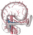
The celiac artery and its branches; the stomach has been raised and the peritoneum removed.
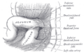
Superior and inferior duodenal fossæ.
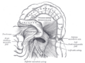
Duodenojejunal fossa.
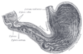
Interior of the stomach.
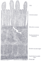
Section of duodenum of cat. X 60.
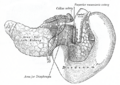
The pancreas and duodenum from behind.
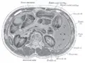
Transverse section through the middle of the first lumbar vertebra, showing the relations of the pancreas.
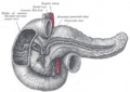
The pancreatic duct.
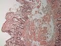
Duodenum with amyloid deposition in lamina propria.
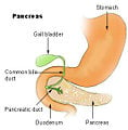
Region of pancreas
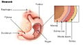
Stomach
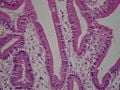
Duodenum with brush border (microvillus)


ReferencesISBN links support NWE through referral fees
- Bender, D. A., and A. E. Bender. 2005. A Dictionary of Food and Nutrition. New York: Oxford University Press. ISBN 0198609612.
- Judge, S. 2001. Duodenum. In C. Blakemore and S. Jennett, The Oxford Companion to the Body. New York: Oxford University Press. ISBN 019852403X.
- Palaeos. 2003. Insectivora. Palaeos. Retrieved July 1, 2007.
- Vetter, R. D., M. C. Carey, and J. S. Patton. 1985. Coassimilation of dietary fat and benzo(a)pyrene in the small intestine: An absorption model using the killifish. Journal of Lipid Research 26: 428-434.
| Digestive system - edit |
|---|
| Mouth | Pharynx | Esophagus | Stomach | Pancreas | Gallbladder | Liver | Small intestine (duodenum, jejunum, ileum) | Colon | Cecum | Rectum | Anus |
Credits
New World Encyclopedia writers and editors rewrote and completed the Wikipedia article in accordance with New World Encyclopedia standards. This article abides by terms of the Creative Commons CC-by-sa 3.0 License (CC-by-sa), which may be used and disseminated with proper attribution. Credit is due under the terms of this license that can reference both the New World Encyclopedia contributors and the selfless volunteer contributors of the Wikimedia Foundation. To cite this article click here for a list of acceptable citing formats.The history of earlier contributions by wikipedians is accessible to researchers here:
The history of this article since it was imported to New World Encyclopedia:
Note: Some restrictions may apply to use of individual images which are separately licensed.
- ↑ Physiology at MCG 6/6ch2/s6ch2_30 - Retrieved December 17, 2007.